Not every bacterial mixture will grow as a biofilm, and that distinction matters a lot. The mixtures most likely to form biofilms are those containing species with strong adhesion proteins (like Staphylococcus aureus or Pseudomonas aeruginosa), combined with partner species that contribute complementary nutrients or structural matrix components, all on a suitable moist surface with available nutrients. If your mixture lacks a solid adherent surface, sufficient moisture, or contains metabolically incompatible species that compete rather than cooperate, biofilm formation is unlikely regardless of how many species are present.
Which Bacterial Mixtures Form Biofilms and Why

Marcus Holloway
31 May 2026
What 'biofilm-forming' actually means in a mixed culture
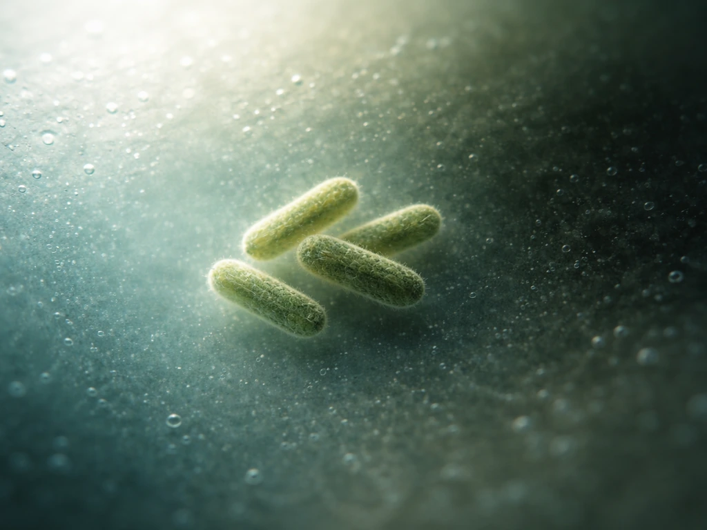
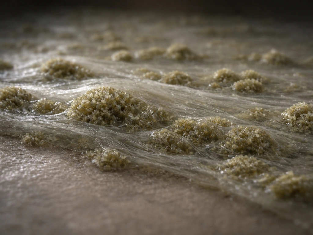
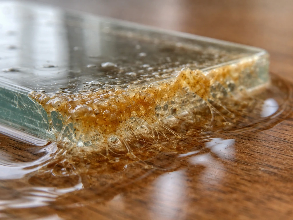
A biofilm is not just bacteria clumping together. The operational definition most researchers use is a matrix-enclosed bacterial population adherent to each other and/or to surfaces or interfaces. Alterations in c-di-GMP turnover proteins can modulate biofilm formation behaviors in Escherichia coli, illustrating how specific regulatory systems influence the capacity to form matrix-enclosed biofilms blank" rel="noopener noreferrer">operational definition most researchers use. That second part, the extracellular polymeric substance (EPS) matrix, is the key. EPS is a mesh of polysaccharides, proteins, nucleic acids, and lipids that bacteria actively secrete. It anchors the community to a surface and protects cells from antibiotics, immune responses, and desiccation. blank" rel="noopener noreferrer">Planktonic (free-floating) bacteria, by contrast, do not produce this matrix and behave completely differently in terms of gene expression, growth rate, and vulnerability.
In a mixed culture, 'biofilm-forming' means at least some species in the community are transitioning from planktonic to the sessile, matrix-producing state. The tricky part is that one strong biofilm-former in a mixture can sometimes scaffold a biofilm that recruits weaker or even non-biofilm-forming species. This is why mixed communities are often more robust biofilm producers than single-species cultures, but it also means you cannot simply assume that because your mixture contains one known biofilm-former, the whole mixture will act accordingly.
Core conditions that every biofilm needs: surface, moisture, and nutrients
Think of biofilm formation as requiring three pillars that must all be present at once. Remove any one of them and the process stalls, regardless of how capable the bacteria are.
A surface to attach to
Bacteria do not biofilm in mid-air or in well-shaken liquid broth. They need an interface: glass, plastic, metal, tissue, teeth, catheter tubing, a pipe wall, food contact surfaces. Even naturally rough or porous materials like wood or unglazed ceramic are especially hospitable because they offer more attachment points. This helps explain why bacteria can colonize agar plates during culture bacteria grow on agar. Surface chemistry matters too. Hydrophobic surfaces (plastics like polystyrene) tend to encourage initial attachment from hydrophobic bacteria, while hydrophilic surfaces may favor different species. This is why the same mixture placed on a polypropylene surface versus a steel surface can produce very different biofilm outcomes.
Moisture
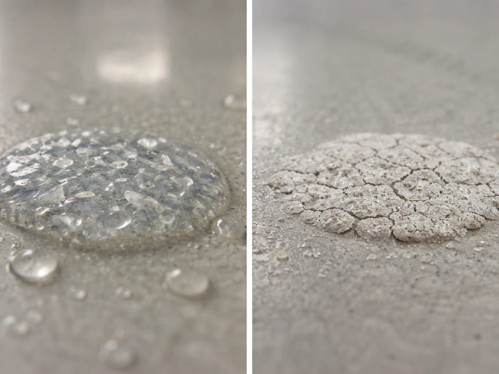
Biofilms absolutely require water activity. The EPS matrix itself is largely water by mass, and bacteria need aqueous conditions both to move toward a surface and to sustain the chemical signaling (quorum sensing) that coordinates matrix production. Environments with water activity below about 0.90 will strongly inhibit most biofilm formation. This is why dry environments, properly dehydrated foods, and desiccated surfaces are much lower risk for biofilm accumulation than wet ones, like sink drains, dental units, or the moist surfaces of respiratory epithelium.
Nutrients
Bacteria forming a biofilm are investing significant metabolic energy in making EPS. They will only commit to this if nutrients are available. Oligotrophic (very low nutrient) conditions tend to keep bacteria in a planktonic, foraging state rather than a sessile, biofilm state. In practice, nutrient-rich interfaces like blood vessel walls, urinary catheters with protein deposits, dental plaque zones, or kitchen drain biofilms are prime biofilm formation sites. Blood agar is also a common growth medium in microbiology labs for culturing and identifying bacteria that may form biofilms what bacteria grow on blood agar. In the lab, using a rich broth (tryptic soy broth, LB broth) will encourage more biofilm than a minimal salts medium for most species.
Oxygen and metabolic compatibility in bacterial mixtures
One of the most common reasons a mixed bacterial inoculum fails to form a biofilm is metabolic incompatibility, and oxygen requirements are the biggest culprit. Obligate aerobes need oxygen to survive; obligate anaerobes are killed by it. Mixing the two and expecting a unified biofilm is unrealistic unless you have a very specific gradient environment, like the deep layers of a thick dental plaque where oxygen is depleted and anaerobes thrive while aerobes colonize the surface. That layered structure is itself a form of metabolic cooperation, but it requires controlled oxygen gradients rather than a well-oxygenated flask.
Facultative anaerobes, species that can use oxygen when it is available but switch to fermentation when it is not, are the most flexible partners in mixed biofilms. Common examples include Escherichia coli, Staphylococcus epidermidis, and many Enterococcus species. Mixtures dominated by facultative anaerobes will biofilm across a wider range of oxygen conditions than mixtures requiring strict aerobic or anaerobic conditions. pH compatibility matters in the same way: most biofilm-forming bacteria operate best between pH 6 and 8, and a mixture where one species acidifies the environment to pH 4.5 through fermentation may inhibit its partners enough to collapse the consortium.
How species traits and surface chemistry influence whether a mixture biofilms
Not all species are equally capable biofilm formers, and the genetic toolkit a species carries determines whether it can anchor to a surface and build matrix. Here are the traits that predict strong biofilm formation:
- Surface adhesins: proteins like FimH (E. coli), IcaA/IcaD (Staphylococcus), or Pel/Psl (Pseudomonas) that physically grab onto surfaces or neighboring cells
- EPS matrix genes: operons encoding polysaccharide synthesis (alginate in Pseudomonas, cellulose in Salmonella and E. coli, PNAG in Staphylococcus)
- Quorum sensing systems: chemical communication that coordinates the switch from planktonic to biofilm mode (e.g., acyl-homoserine lactone signaling in Gram-negatives, autoinducer-2 in mixed communities)
- Motility with surface-sensing: flagella that allow initial approach to a surface, then trigger down-regulation once contact is made
- c-di-GMP regulation: the intracellular second messenger that in high concentrations drives biofilm formation and at low concentrations promotes planktonic dispersal
When you know the Gram stain category of your mixture, that alone gives you useful predictions. Gram-negative rods like Pseudomonas aeruginosa, E. coli, Klebsiella pneumoniae, and Acinetobacter baumannii are notorious biofilm formers, especially on abiotic (non-living) surfaces. Gram-positive cocci like Staphylococcus aureus and Staphylococcus epidermidis are among the most clinically significant biofilm producers on implanted devices. Some Gram-positives like Bacillus subtilis form architecturally sophisticated biofilms on solid surfaces. By contrast, some species, many Campylobacter strains, certain Mycoplasma species, and many obligate intracellular pathogens, are poor or non-biofilm formers.
| Bacterial group / genus | Biofilm tendency | Key trait driving formation | Common habitat |
|---|---|---|---|
| Pseudomonas aeruginosa | Very high | Pel/Psl polysaccharides, alginate, quorum sensing | Wounds, lungs, water systems |
| Staphylococcus aureus / epidermidis | Very high | PNAG (icaADBC operon), adhesins | Skin, catheters, implants |
| Escherichia coli (uropathogenic) | High | Curli fibers, cellulose, type 1 fimbriae | Urinary tract, gut, surfaces |
| Klebsiella pneumoniae | High | Capsular polysaccharides, fimbriae | Urinary tract, respiratory |
| Bacillus subtilis | High (model organism) | Sporulation-linked, TasA protein matrix | Soil, plant roots, lab surfaces |
| Streptococcus mutans | High (dental specific) | Glucans from sucrose, adhesins | Dental plaque |
| Enterococcus faecalis | Moderate to high | Esp surface protein, aggregation substance | GI tract, urinary tract, wounds |
| Salmonella enterica | Moderate | Curli, cellulose production | Food-contact surfaces, gut |
| Lactobacillus spp. | Variable (strain-dependent) | Surface proteins, EPS | Gut, vaginal mucosa |
| Campylobacter jejuni | Low to moderate | Limited adhesins, microaerophilic restrictions | GI tract (mostly planktonic) |
Mixed cultures that combine a high-biofilm-tendency species with a metabolically complementary partner are the most predictable biofilm formers. Classic examples include Pseudomonas aeruginosa with Staphylococcus aureus in wound infections, and Streptococcus gordonii with Porphyromonas gingivalis in periodontal disease. In particular, researchers often ask on which type of media Streptococcus can grow when planning experiments or interpreting biofilm results. In both cases, early colonizers modify the surface or produce signaling molecules that recruit the secondary species.
Practical ways to predict or screen biofilm formation today
If you are working with a specific mixture and need to know whether it biofilms, here is a practical screening workflow you can run with standard microbiology equipment.
- Identify your species: Before anything else, know what is in your mixture, even at the Gram class level. Use Gram staining, selective media, or 16S rRNA sequencing data if available. Cross-reference species against known biofilm-forming genera using published literature or databases like NCBI or the BioFilm Database (BIOFILM-Q).
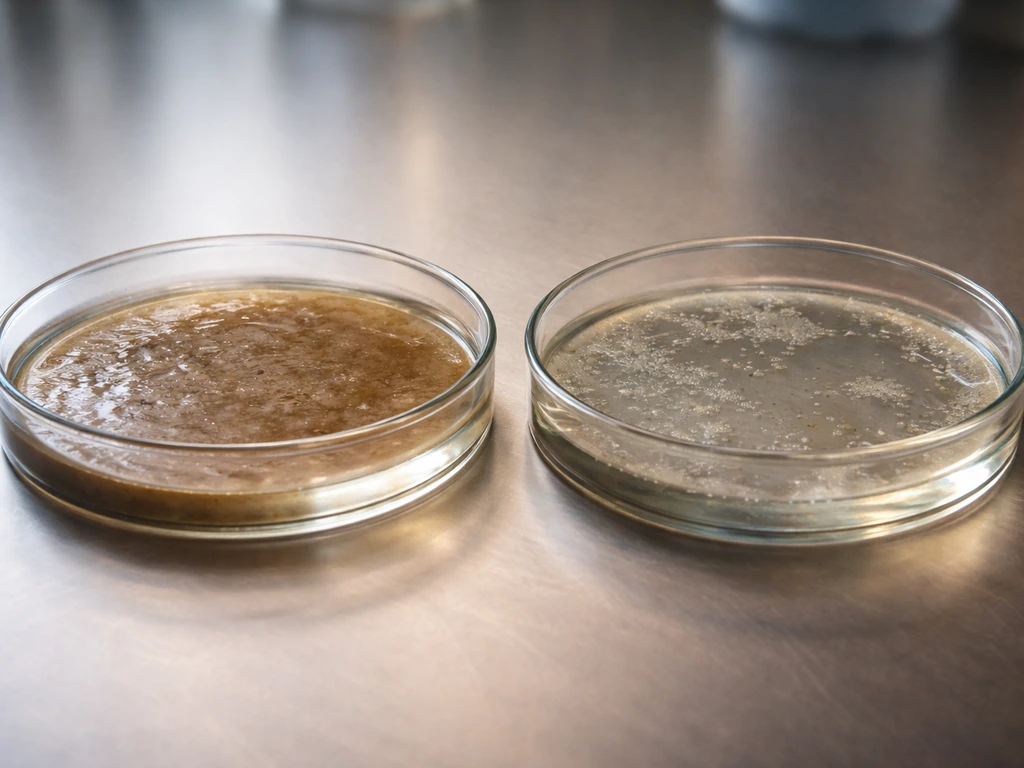
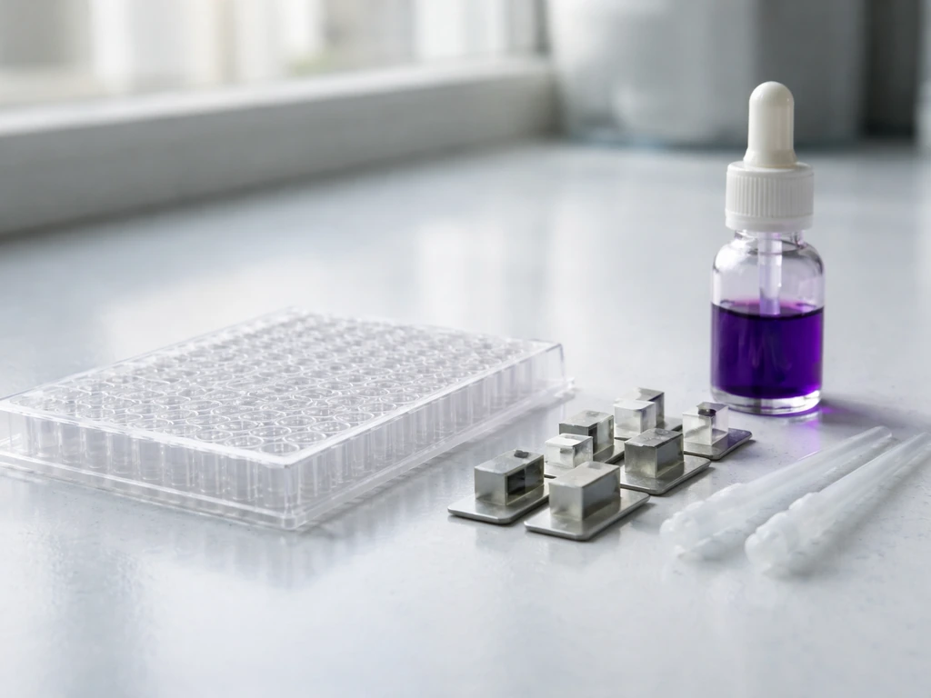
- Choose the right surface: Use 96-well polystyrene microtiter plates for high-throughput screening. Polystyrene is the standard surface for the crystal violet (CV) biofilm assay because it is hydrophobic and promotes initial adhesion for many clinically relevant species.
- Set up the crystal violet assay: Inoculate wells with your bacterial mixture in a nutrient-rich broth (tryptic soy broth works for most mixtures). Incubate statically (no shaking) at the appropriate temperature, typically 37°C for clinical isolates, for 24 to 48 hours. After incubation, remove planktonic cells by washing gently with phosphate-buffered saline. Fix adherent cells with methanol, stain with 0.1% crystal violet for 15 minutes, then wash again. Dissolve the stain with 95% ethanol and read absorbance at 595 nm. Higher absorbance means more biofilm biomass.
- Interpret absorbance values: An OD595 ratio of the stained biofilm to a sterile control greater than 2 is generally considered a positive biofilm result. Values between 1 and 2 suggest weak biofilm formation; below 1 is typically negative.
- Visual and microscopic confirmation: Look for a visible ring or pellicle at the air-liquid interface in test tubes, or a cloudy film on the well bottom under a dissecting microscope. For more detail, use confocal laser scanning microscopy with live/dead staining (SYTO9 and propidium iodide) to visualize the 3D architecture and cell viability within the biofilm.
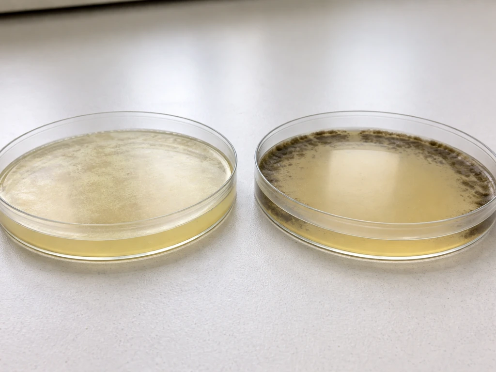
- Check for EPS production: If you have access to scanning electron microscopy (SEM), biofilms show characteristic interconnected cells embedded in matrix. Light-level alternatives include staining with Congo red agar (CRA); species producing EPS form black or dark red colonies, while non-producers remain pink or red.
- Run positive and negative controls: Always include a known strong biofilm-former (P. aeruginosa PAO1 or S. epidermidis RP62A) as a positive control and a biofilm-negative strain (like E. coli MG1655 fimH deletion mutant) as a negative control.
For students and classroom settings without access to spectrophotometers, a simplified version of this screen uses visual scoring of pellicle formation in glass tubes and Congo red agar plates, which can give a reliable qualitative answer about whether a mixture contains biofilm-forming members. If you are trying to answer what agar Staphylococcus aureus grows on, picking the right media matters as much as the basic biofilm screen Congo red agar plates.
Common reasons mixtures fail to form biofilms, and how to fix them
If your mixture is not forming a biofilm when you expect it to, the problem is almost always one of these fixable conditions:
| Problem | Why it prevents biofilm | How to fix it |
|---|---|---|
| Shaking or agitation during incubation | Disrupts initial surface attachment before cells can anchor | Use static incubation for the first 24 hours |
| Too little inoculum | Quorum sensing signals do not reach threshold for biofilm gene expression | Increase starting inoculum to OD600 0.05 to 0.1 in the well |
| Wrong incubation temperature | Optimal adhesion gene expression is temperature-dependent (e.g., 37°C for clinical isolates) | Match temperature to the species' natural host environment |
| Nutrient-poor medium | Insufficient energy for EPS matrix production | Switch to TSB, LB, or BHI broth instead of minimal media |
| Incompatible species pairing | One species produces bacteriocins or metabolic byproducts that kill the other | Test species individually first; mix only those that are individually viable together |
| Surface too hydrophilic for the species | Poor initial adhesion | Test on multiple surfaces (glass vs. polystyrene vs. silicone) |
| Short incubation time | Biofilm architecture takes 24 to 72 hours to fully develop | Extend incubation; check at 24, 48, and 72 hours |
| Wrong pH | Extreme pH inhibits signaling and EPS production | Buffer medium to pH 7.0 to 7.4 unless testing pH-specific environments |
A point worth emphasizing: competition between species is one of the most underappreciated reasons mixed cultures fail to biofilm. If a fast-growing species like E. coli outcompetes a slower but stronger biofilm-former like P. aeruginosa in the early growth phase, the mixture may look like a simple planktonic culture. Adjusting the inoculum ratio or pre-growing each species separately before mixing can help reveal the true biofilm potential of the community.
Safety, limitations, and how to interpret your results
Several of the bacterial genera most relevant to biofilm work, Pseudomonas aeruginosa, Staphylococcus aureus, Klebsiella pneumoniae, and uropathogenic E. coli - are BSL-2 organisms. Work with these species requires appropriate containment: lab coat, gloves, biosafety cabinet for aerosol-generating steps, and proper waste decontamination (autoclaving liquid waste before disposal). Never assume a clinical or environmental isolate is harmless because it is commonly studied.
Results from in vitro biofilm assays have real limitations you need to keep in mind. A biofilm that forms in a polystyrene well under static broth conditions may behave very differently on living tissue, under flow conditions (like a urinary catheter with urine flow), or in the presence of host immune factors. The crystal violet assay measures total biomass including dead cells and matrix, not just living biofilm-forming cells, so high absorbance does not automatically mean a metabolically active, clinically significant biofilm. Pair quantitative assays with visual confirmation and, when possible, viable cell counts from the scraped biofilm.
It is also worth noting that biofilm formation is a strain-level trait, not just a species-level one. Two strains of E. coli can behave completely differently: one may be a robust biofilm former due to curli fiber and cellulose production, while another (especially laboratory-adapted strains) may have lost those traits entirely. This is why published results for a species do not always predict what your specific isolate will do. Always validate your actual strain rather than assuming behavior from the literature.
Finally, interpreting mixed-species results requires care. A positive biofilm signal from a mixture tells you that at least one species is forming a biofilm, but not necessarily which one. To identify which species in the mixture are the primary biofilm contributors, you need to test each species individually and then in pairwise or higher-order combinations. This combinatorial approach is more work, but it is the only way to truly answer which components of a bacterial mixture are growing as a biofilm rather than just riding along in one.
FAQ
If my bacterial mixture forms a biofilm, does that mean all species in the mix are growing as biofilms?
Not necessarily. A mixture can produce a biofilm signal even if only one member is matrix-producing, with other species passively attached inside the EPS. To determine which organisms are actually biofilm growers, test each species alone and in pairwise combinations, then compare matrix staining and viable counts rather than relying on total biomass alone.
Can obligate aerobes and obligate anaerobes in the same mixture ever form a single biofilm together?
Yes, especially if oxygen and nutrient gradients form naturally. Obligate aerobes and obligate anaerobes usually cannot coexist in a single uniform environment, but a thick surface layer (for example, dense plaque or a biofilm on a rough porous material) can create low-oxygen pockets where anaerobes survive while aerobes persist at the outer edge.
Why does the same bacterial mixture biofilm under static conditions but not in shaking broth?
Use the interface conditions to your advantage. Static, nutrient-rich liquid with a surface tends to promote attachment and EPS production, while shaking disrupts the initial “settling” step and can prevent stable EPS anchoring. If you want to know what in your mixture truly enables biofilm growth, compare static versus gently mixed setups with the same inoculum and surface type.
How can I tell whether the mixture is producing living biofilm versus just accumulating dead cells and matrix?
Many “biofilm-positive” results are due to biomass, not living, matrix-active growth. Congo red, crystal violet, and similar assays can overcount dead cells and nonviable matrix. To confirm that bacteria are truly growing in the biofilm state, pair staining with viable recovery from scraped biofilm (for example, plate counts) and, if available, metabolic readouts.
If the article says a genus or species is a biofilm former, why might my isolate still fail to form a biofilm?
Strain differences are a common reason. Even within the same species, one strain may express curli fibers, cellulose, or strong adhesion factors, while another may have lost key genes or regulatory pathways. Always test your exact isolates, and do not assume “species X biofilms” will match “my strain X biofilms.”
What nutrient conditions are most likely to switch bacteria from planktonic growth into biofilm growth?
Nutrient availability alone can be misleading. Very low nutrients often keep cells in a foraging, planktonic state, but extremely high nutrients can also shift behavior toward faster planktonic growth depending on the species and surface. A practical next step is to test a small nutrient gradient (for example, low, medium, high) at the same moisture level and surface chemistry.
What should I change if my mixed culture fails to form a biofilm even though one member is known to be a strong biofilm former?
Competition is often strongest during early attachment. If a fast grower dominates the first hours, it may prevent the primary biofilm former from establishing enough surface contact to initiate EPS scaffolding. Try adjusting inoculum ratios and, when feasible, pre-grow each species separately and mix at a timing that allows the intended “early colonizer” to attach first.
Will a mixture that forms a biofilm in a lab well also form biofilms in real flow environments?
Yes. Flow typically changes whether bacteria can remain attached long enough to produce and mature EPS. For catheter-like or pipe-wall relevance, include flow or shear conditions, because a mix that biofilms in a polystyrene well under static broth may disperse under urine-like flow.
How do surface materials change which bacterial mixture members end up growing as a biofilm?
Hydrophobicity can affect which member attaches first, which then determines who gets recruited into the community. If your goal is to see which mixture components can grow as biofilms, test multiple common surfaces (for example, hydrophobic plastic and steel/glass) using the same inoculum and moisture, because “biofilm-ready” may mean “surface-compatible.”
What does a positive mixed-culture biofilm test actually tell me, and what is the next step to identify the main biofilm growers?
A positive result means at least one species can transition to a matrix-producing sessile state under your conditions, but it does not identify the primary contributor. To answer “which bacteria are growing as a biofilm,” run individual and combinatorial tests, then use selective confirmation (for example, species-specific recovery or targeted identification) on the scraped biofilm.