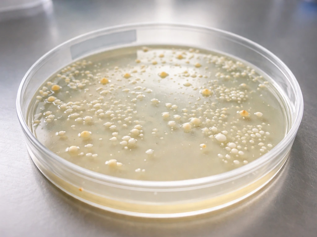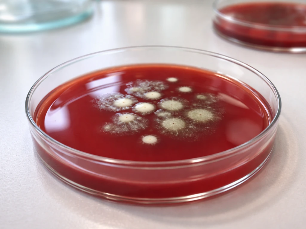
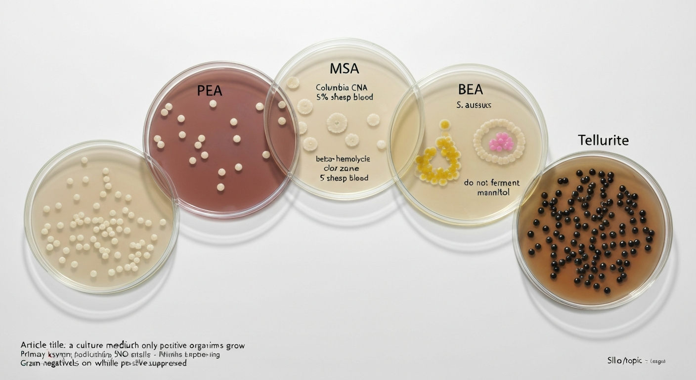
A culture medium on which only Gram-positive organisms grow is called a selective medium, and the classic examples include Phenylethyl Alcohol (PEA) agar, Columbia CNA agar (Colistin-Nalidixic Acid), Mannitol Salt Agar (MSA), Bile Esculin Agar (BEA), and tellurite-based media. Each one contains one or more chemical agents, high salt, specific antibiotics, bile salts, or toxic compounds like tellurite, that physically or biochemically block Gram-negative bacteria from establishing colonies, while leaving most Gram-positive organisms free to grow. Understanding why that works requires looking at what makes Gram-positive and Gram-negative cell walls so different, and why that difference is their undoing when the wrong chemicals show up in the agar.
A culture medium on which only Gram-positive organisms grow.

What Gram-positive and Gram-negative actually mean
The Gram stain, developed by Hans Christian Gram in 1884, separates bacteria into two broad categories based on how they respond to a crystal violet and iodine staining procedure. Gram-positive bacteria have a thick peptidoglycan cell wall (roughly 20–80 nm) that traps the purple crystal violet-iodine complex inside the cell, so they appear purple or violet under a microscope. Gram-negative bacteria have a much thinner peptidoglycan layer but add a second, outer membrane made largely of lipopolysaccharide (LPS). That outer membrane flushes the crystal violet out during the decolorization step, and the cells pick up a pink or red counterstain instead. This structural difference is not just a lab curiosity, it has enormous consequences for how each group responds to antibiotics, disinfectants, and the selective ingredients in culture media.
When we say a medium allows 'only' Gram-positive organisms to grow, we mean it strongly suppresses the growth of Gram-negative species under standard incubation conditions. In practice, a small number of unusually resistant Gram-negative strains or highly salt-tolerant species may still produce occasional colonies, which is why microbiologists describe growth on selective media as 'presumptive' rather than definitive. Selectivity is a matter of degree, not an absolute wall.
Selective versus differential media: two different jobs
Students often conflate selective and differential media because many classic plates are actually both at the same time. It helps to separate the concepts clearly before combining them.
A selective medium contains agents that inhibit certain organisms while permitting others to grow. The goal is to narrow down who shows up on the plate. Selectivity does not tell you anything about what the surviving organism is doing metabolically, it just controls who gets a seat at the table.
A differential medium, by contrast, includes indicators (typically pH-sensitive dyes, iron salts, or chromogenic substrates) that produce visible color changes when an organism carries out a specific metabolic reaction, like fermenting a sugar or hydrolyzing a compound. Differential features help you distinguish between organisms that can both grow on the same medium.
Mannitol Salt Agar is a good teaching example of a medium that is both: the high salt concentration is the selective element (it suppresses most Gram-negatives and many Gram-positives), while the mannitol and phenol red indicator are the differential elements (acid production from mannitol fermentation turns the medium yellow). MacConkey agar flips the situation, it is selective against Gram-positives but differential among Gram-negatives based on lactose fermentation. Keeping this distinction in mind helps you read plates and interpret results correctly.
How selective media block Gram-negative bacteria
The outer membrane of Gram-negative bacteria is their defining structural feature and, ironically, their vulnerability when specific selective agents are present. Here is how the most common mechanisms work:
Bile salts and high pH
Bile salts (sodium deoxycholate, sodium taurocholate) are detergent-like molecules that disrupt lipid bilayers. Gram-negative bacteria have an outer membrane rich in phospholipids, which bile salts attack directly. Gram-positive bacteria lack this outer membrane, and their thick peptidoglycan wall provides a degree of resistance to bile. Media like MacConkey agar use bile salts to exclude Gram-positives, while Bile Esculin Agar uses a lower bile concentration specifically to exclude most Gram-positives while allowing bile-tolerant Gram-positives, primarily Enterococcus and group D streptococci, to survive.
High sodium chloride (NaCl)
Mannitol Salt Agar contains approximately 7.5 to 10% NaCl, creating a hypertonic environment that is lethal to most bacteria. Most Gram-negative species are osmotically sensitive and cannot survive at these salt concentrations. Staphylococcus species are halotolerant (salt-tolerant) and thrive under these conditions, which is why MSA is used to selectively recover them from mixed-flora specimens. Think of it like putting a plant in saltwater, most cells shrink and die through osmosis, but a select few have mechanisms to balance the osmotic pressure and keep growing.
Phenylethyl alcohol (PEA)
Phenylethyl alcohol works by altering membrane permeability in susceptible Gram-negative bacteria and interfering with DNA synthesis. Because Gram-negative cells have that lipid-rich outer membrane, PEA has easier access to disrupt membrane integrity and block replication. Gram-positive organisms are far less affected at the concentrations used in PEA agar (typically around 0.25%). PEA agar is often supplemented with 5% sheep blood to enrich the medium and support the growth of fastidious Gram-positive organisms like streptococci.
Colistin and nalidixic acid (CNA)
CNA agar uses a two-antibiotic combination that hits Gram-negatives from two different angles. Colistin (a polymyxin antibiotic) binds directly to lipid A, the lipopolysaccharide component of the Gram-negative outer membrane, causing membrane disruption and cell death. Nalidixic acid, an early quinolone, inhibits DNA gyrase and topoisomerase, blocking DNA replication. Together, these two agents create a highly effective chemical barrier against Gram-negative bacilli at the concentrations used commercially (typically around 10 mg/L each). Gram-positive bacteria largely lack the outer membrane target for colistin and are intrinsically less susceptible to nalidixic acid at these concentrations, so they grow undisturbed on the enriched Columbia blood agar base.
Potassium tellurite
Tellurite (TeO3^2-) is toxic to most bacteria because it generates reactive oxygen species (ROS) inside cells and disrupts thiol chemistry critical for cellular function. Most bacteria cannot survive exposure to tellurite. Certain Gram-positive species, most famously Corynebacterium diphtheriae, as well as some Staphylococcus and Bacillus strains, possess enzymatic reductase systems and thiol-based detoxification pathways that reduce tellurite to elemental tellurium (Te0), which deposits as a visible black precipitate in and around the colony. This reduction is a detoxification mechanism, and the resulting black colonies are a classic diagnostic clue. Genetic determinants like the tehAB operon have been linked to tellurite resistance, though the full picture is still being researched.
The main selective media that favor Gram-positive organisms
Phenylethyl Alcohol (PEA) Agar
PEA agar is one of the cleanest selective media available for Gram-positive recovery. It inhibits Gram-negative bacilli (particularly swarming Proteus species, which would otherwise overgrow a plate) and supports the growth of Gram-positive cocci and rods. The blood-supplemented version supports fastidious organisms like streptococci and allows hemolysis patterns to be observed. It is especially useful in clinical settings when a specimen like a wound swab contains a mixed population of organisms.
Columbia CNA Agar
CNA agar is probably the most commonly used selective medium for Gram-positive organisms in clinical microbiology. Its enriched Columbia base supports fastidious Gram-positive organisms, while colistin and nalidixic acid suppress most Gram-negative rods. The addition of 5% sheep blood allows hemolysis evaluation, critical for differentiating streptococcal species. Because it supports a wide range of Gram-positive organisms without requiring unusual conditions, CNA is often used in combination with a Gram-negative selective medium (like MacConkey) when plating mixed clinical specimens.
Mannitol Salt Agar (MSA)
MSA is the workhorse medium for Staphylococcus identification, both in clinical labs and food microbiology. The 7.5 to 10% NaCl selects for halotolerant organisms, and the mannitol plus phenol red combination differentiates mannitol fermenters (acid production turns the medium yellow, strongly associated with S. aureus) from non-fermenters (medium stays red or pink). It is worth noting that MSA selectivity is not absolute: Enterococcus, Micrococcus, and some Bacillus species can also grow, and occasional salt-tolerant Gram-negative isolates have been reported. Enterococcus species are halotolerant and can grow on high‑salt media such as MSA (Enterococcus, NCBI Bookshelf, taxonomic/physiologic notes on salt tolerance) Enterococcus — NCBI Bookshelf (taxonomic/physiologic notes; salt tolerance). Similarly, some S. aureus strains with defective mannitol utilization genes will not turn the medium yellow, so MSA results are always considered presumptive.
Bile Esculin Agar (BEA)
Bile Esculin Agar is both selective and differential, designed specifically to identify Enterococcus species and group D streptococci. The bile concentration (typically 40%) inhibits most Gram-positive organisms except those that are bile-tolerant. Organisms that survive and also hydrolyze esculin produce esculetin, which reacts with ferric ions (from ferric citrate in the medium) to form a black or dark brown precipitate around the colony. Studies report sensitivity above 99% and specificity around 97% for Enterococcus identification using standardized 40% bile conditions with 24-hour incubation, but performance depends heavily on the exact bile concentration, inoculum size, and incubation time used.
Tellurite-based media
Tellurite media include formulations like Hoyle's medium and Cystine-Tellurite Blood Agar (CTBA), historically used for isolating Corynebacterium diphtheriae (the diphtheria pathogen). The black colony appearance from tellurite reduction is diagnostically useful. Tellurite-containing plates are also used in some Staphylococcus isolation protocols. Because tellurite is broadly toxic, these media tend to be more selective than PEA or CNA, but they are also more demanding on fragile or fastidious Gram-positive species, so they are used for specific clinical contexts rather than broad Gram-positive recovery.
Which Gram-positive organisms actually grow on these media
Not every Gram-positive organism grows equally well on every selective medium, and this is a common source of confusion. Here is a breakdown of the major players:
Staphylococcus aureus
S. aureus is arguably the organism most associated with Gram-positive selective media. It grows well on PEA, CNA, and MSA. For more details on preferred media and comparative growth characteristics, see what agar does Staphylococcus aureus grow on. On MSA, most strains produce yellow colonies due to mannitol fermentation, making them immediately distinguishable from other staphylococci. On CNA with blood, S. aureus typically produces beta-hemolysis (complete clearing of red blood cells around the colony). Its halotolerance, mannitol fermentation capacity, and resistance to colistin and nalidixic acid all reflect its robust physiology, features that also make it a clinically important pathogen in wound infections, food poisoning, and hospital-acquired infections.
Other Staphylococcus species
Coagulase-negative staphylococci (CoNS), such as Staphylococcus epidermidis and Staphylococcus saprophyticus, also grow on PEA, CNA, and MSA. On MSA, they typically produce pink or red colonies (no mannitol fermentation), which helps distinguish them from S. aureus presumptively. CoNS are increasingly important clinically as causes of infections in immunocompromised patients and device-associated infections.
Streptococcus species
Streptococci grow well on PEA agar and CNA agar when blood is added, because they are fastidious, they need enriched nutrients and do not grow reliably on minimal media. See on which type of media will Streptococcus grow for a focused list of media that support streptococcal growth. The blood supplement allows you to observe hemolysis patterns: beta-hemolytic streptococci (like Streptococcus pyogenes, group A strep) produce complete clearing, alpha-hemolytic species (like Streptococcus pneumoniae) produce partial greenish clearing, and gamma-hemolytic (non-hemolytic) species show no change. Streptococci generally do not grow on MSA, which is one reason MSA is so useful for Staphylococcus-specific recovery.
Enterococcus
Enterococcus faecalis and Enterococcus faecium are robust Gram-positive organisms that grow on PEA, CNA, MSA (due to halotolerance), and are the signature organisms of Bile Esculin Agar. Their ability to survive bile, hydrolyze esculin, and tolerate high salt concentrations reflects their natural habitat in the gastrointestinal tract. Enterococci are important in food safety contexts, their presence in certain foods can indicate fecal contamination, and they are significant clinical pathogens due to rising antibiotic resistance.
Non-selective media: why blood agar and nutrient agar grow almost everything
Blood agar (typically 5% sheep blood on a tryptic soy or Columbia agar base) is the classic non-selective, enriched medium in clinical microbiology. It contains no selective agents, no antibiotics, no bile, no excess salt. The blood provides hemin, NAD, and other growth factors that support even the most fastidious organisms. As a result, both Gram-positive and Gram-negative organisms grow freely on blood agar, and the plate's main diagnostic value comes from the hemolysis patterns rather than from any restriction on who can grow. For more background on the basic reasons why bacteria grow on agar, see why does bacteria grow on agar. For a concise list of organisms commonly recovered on blood agar, see what bacteria grow on blood agar. In fact, most Gram-positive and Gram-negative organisms will grow on blood agar, making it a broadly permissive non-selective medium. This is a key contrast with selective media: blood agar is an open invitation to everyone in the sample.
Nutrient agar is even simpler, it contains beef extract, peptone, and agar, providing a basic nutritional scaffold with no enrichment and no selectivity. It supports the growth of most non-fastidious Gram-positive and Gram-negative organisms, making it ideal for teaching general bacterial growth principles and for environmental sampling where you want to know the total viable count rather than just one group. Neither blood agar nor nutrient agar can be used to confirm that an isolate is Gram-positive; for that, you need either the Gram stain itself or a selective medium.
This is why the combination approach matters in a real laboratory: plating a clinical specimen simultaneously onto CNA (for Gram-positives), MacConkey (for Gram-negative enteric bacilli), and blood agar (for everything, especially fastidious organisms) gives a much more complete picture than any single plate alone.
Practical applications: classroom, food safety, and clinical settings
In a microbiology classroom, selective media are powerful teaching tools because they make an abstract concept, 'this organism has this property', visible as a color change or a clear zone on a plate. MSA is a favorite because students can observe the yellow color change from S. aureus mannitol fermentation in real time, connecting biochemistry to the physical appearance of a colony. This kind of hands-on observation is one of the best ways to internalize the difference between selective and differential features.
In food safety, MSA is used to detect Staphylococcus aureus in foods because S. aureus produces heat-stable enterotoxins responsible for one of the most common forms of food poisoning. The high-salt selectivity of MSA is particularly useful for testing processed foods (like cured meats, cheeses, and ready-to-eat products) that themselves contain elevated salt levels, conditions that would inhibit many competing organisms but not S. aureus. Bile Esculin Agar is similarly used in food microbiology for Enterococcus detection as a hygiene indicator.
In clinical microbiology, selective Gram-positive media are used daily for processing wound swabs, respiratory specimens, urine cultures, and blood culture sub-cultures. A rapid presumptive identification from a selective plate can guide empirical antibiotic therapy decisions before final confirmation results are available, making the choice of media a genuinely consequential clinical decision.
A note on biofilm formation and how media influences it
Most selective Gram-positive media are designed for planktonic (free-floating) colony growth and are not optimized for biofilm detection. Biofilms, structured communities of bacteria encased in a self-produced matrix, form preferentially on surfaces and under specific nutrient and flow conditions that standard agar plates do not replicate well. That said, organisms like Staphylococcus epidermidis and Staphylococcus aureus that are routinely recovered on MSA and CNA are among the most biofilm-competent bacteria known, particularly in device-associated infections. The growth conditions on a selective plate give you a snapshot of who is present, but not necessarily how those organisms are behaving in their natural environment, a reminder that in vitro culture is always a simplified model of real-world microbiology. For more information on which bacterial mixtures grow as biofilms, consult resources on biofilm formation and mixed-species biofilms.
How to interpret results from selective Gram-positive media
A few practical interpretation principles are worth keeping in mind whenever you work with selective Gram-positive media:
- Growth on a selective Gram-positive medium is presumptive, not definitive. It tells you the organism is likely Gram-positive and has the relevant tolerance (salt, bile, etc.), but it does not give you a species identification.
- Confirmatory testing — Gram stain, biochemical panels, MALDI-TOF mass spectrometry, or molecular methods — is always required for final organism identification in clinical and food safety contexts.
- Absence of growth on a selective medium does not guarantee the organism is absent from the specimen. A very low inoculum, a fastidious strain, or inhibition by competing organisms can all cause false negatives.
- Color changes on differential media (yellow on MSA, black precipitate on BEA) are strong presumptive indicators but have documented exceptions — some S. aureus strains do not ferment mannitol, and some non-aureus species can.
- Incubation time matters: most selective media are read at 18 to 24 hours, but some fastidious organisms may require 48 hours. Reading plates too early can miss slow-growing organisms.
- Quality control (QC) with known positive and negative control organisms is a professional standard (per CLSI guidelines) that ensures media are performing as expected before patient or food samples are processed.
Limitations of selective media and important safety notes
No selective medium is perfect. The categories 'Gram-positive only' and 'Gram-negative only' are useful approximations, not absolute rules. Unusual organisms, antibiotic-resistant strains, and environmental isolates can break the expected patterns. For example, some Gram-negative organisms with acquired salt tolerance or antibiotic resistance may grow on media where they would normally be suppressed. Conversely, some Gram-positive organisms are sensitive to the selective agents in a given medium, certain streptococcal species grow poorly on MSA, and some Gram-positive rods are inhibited by high PEA concentrations.
From a safety standpoint, working with selective media for known or suspected pathogens (S. aureus, C. diphtheriae, Enterococcus with antibiotic resistance) requires appropriate biosafety practices. This article is intended as an educational explanation of the principles behind these media and is not a laboratory protocol. Specific procedural guidance should come from institutional standard operating procedures, manufacturer package inserts, and applicable biosafety guidelines.
Comparing common media at a glance
| Medium | Selective agent(s) | Differential feature | Typical Gram-positive organisms recovered | Gram-negative fate |
|---|---|---|---|---|
| PEA (Phenylethyl Alcohol) Agar | Phenylethyl alcohol (~0.25%) | Hemolysis if blood added | Staphylococcus spp., Streptococcus spp., Enterococcus spp. | Suppressed (esp. Gram-negative rods) |
| CNA (Columbia CNA) Agar | Colistin (~10 mg/L) + nalidixic acid (~10 mg/L) | Hemolysis patterns (alpha, beta, gamma) | Staphylococcus aureus, Streptococcus spp., Enterococcus spp. | Most suppressed by colistin and nalidixic acid |
| Mannitol Salt Agar (MSA) | ~7.5–10% NaCl (high salt) | Mannitol + phenol red: yellow = acid (S. aureus presumptive); red/pink = no fermentation | Staphylococcus aureus (yellow), CoNS (pink/red), Enterococcus spp., Micrococcus spp. | Most suppressed; occasional salt-tolerant exceptions |
| Bile Esculin Agar (BEA) | ~40% bile salts | Esculin hydrolysis + ferric citrate: black precipitate = positive | Enterococcus spp., group D streptococci | Suppressed by bile; most Gram-negatives inhibited |
| Tellurite media (e.g., CTBA, Hoyle's) | Potassium tellurite | Black colonies = tellurite reduction | Corynebacterium diphtheriae, some Staphylococcus and Bacillus spp. | Suppressed; tellurite toxic to most Gram-negatives |
| Blood Agar (5% sheep blood) | None (non-selective) | Hemolysis: alpha (green), beta (clear), gamma (none) | All Gram-positives including fastidious species | Grows freely; non-selective |
| Nutrient Agar | None (non-selective) | None | Non-fastidious Gram-positive organisms | Grows freely; non-selective |
| MacConkey Agar (contrast) | Bile salts + crystal violet | Lactose fermentation: pink/red = lactose fermenter; colorless = non-fermenter | Suppressed (Gram-positive contrast medium) | Gram-negatives recovered selectively |
The table makes one practical point very clear: the same structural feature that defines Gram-negative bacteria (the outer membrane with LPS) is the feature that makes them vulnerable to bile salts, colistin, and PEA. Remove that outer membrane, as you effectively do by working with Gram-positive organisms, and those selective agents lose most of their punch. That is the biological principle underlying every Gram-positive selective medium ever developed, and it is the single idea worth taking away from this entire topic.
FAQ
What does the phrase “a culture medium on which only Gram‑positive organisms grow” mean?
It describes a selective medium formulated to inhibit most Gram‑negative bacteria while permitting many Gram‑positive bacteria to grow. In practice this is presumptive — some Gram‑positives grow and a few Gram‑negatives may tolerate the medium. Such media enrich for Gram‑positive organisms to simplify isolation and presumptive identification, but they do not provide definitive species identification.
How do selective and differential media differ?
Selective media contain agents (salts, bile, antibiotics, dyes, oxidizing agents) that inhibit unwanted groups of microbes and favor others. Differential media contain indicators (pH dyes, precipitating reagents) or substrates that produce visible changes revealing metabolic differences between organisms (for example, fermenters turning an indicator yellow). A single medium can be both selective and differential.
What biochemical or physical mechanisms are used to inhibit Gram‑negative bacteria on these media?
Common mechanisms include: antibiotics that target Gram‑negative features (e.g., colistin/polymyxins bind LPS and disrupt the outer membrane; nalidixic acid inhibits DNA gyrase), high salt concentrations that select for halotolerant organisms, bile salts that solubilize membranes of susceptible bacteria, oxidizing agents or toxic oxyanions (e.g., tellurite) that many cells cannot detoxify. These agents exploit structural or metabolic differences between taxa.
What are concrete examples of media that select for Gram‑positive organisms and how do they work?
- Phenylethyl alcohol (PEA) agar: phenethyl alcohol disrupts membrane function and can interfere with DNA synthesis in many Gram‑negatives, permitting Gram‑positive recovery (often with added blood). - Columbia CNA (colistin‑nalidixic acid) blood agar: colistin disrupts Gram‑negative outer membranes by binding lipid A; nalidixic acid inhibits DNA gyrase — together inhibit many Gram‑negatives. - Mannitol Salt Agar (MSA): high NaCl (~7.5–10%) selects for halotolerant organisms (classically Staphylococcus spp.); mannitol + phenol red provides differential mannitol fermentation. - Bile esculin agar: bile inhibits many Gram‑positives; organisms that hydrolyze esculin produce esculetin which reacts with ferric ions to form a black precipitate (used for Enterococcus/group D streptococci). - Tellurite‑containing media: some Gram‑positives reduce tellurite to elemental tellurium, producing black colonies (used for Corynebacterium and others).
Which clinically important species typically grow on these media?
Common Gram‑positive genera recovered include Staphylococcus spp. (Staphylococcus aureus, S. epidermidis), Streptococcus spp. and Enterococcus spp. (Enterococcus faecalis, E. faecium), Corynebacterium spp., Bacillus spp., and Micrococcus. Growth patterns (e.g., mannitol fermentation, bile‑esculin hydrolysis, hemolysis on blood agar) provide presumptive clues to identity.
Are these media perfectly exclusive to Gram‑positives?
No. Selectivity is relative. Some Gram‑negative strains can tolerate selective agents (salt‑tolerant or antibiotic‑resistant strains), and some non‑target Gram‑positives may be inhibited. Additionally, media like MSA select for halotolerance rather than Gram‑positivity, so other halotolerant taxa (including occasional Gram‑negatives) can grow. Therefore results are presumptive and require confirmatory testing (biochemical assays, MALDI‑TOF, molecular methods).