Both Gram-positive and Gram-negative bacteria can grow on blood agar. The medium does not filter by Gram reaction at all. Blood agar is a general-purpose, enriched medium, meaning it is designed to support a wide range of organisms rather than select for or against any particular group. If a bacterium has basic nutritional needs and tolerates standard incubation conditions (around 35 to 37°C, aerobic or facultative), blood agar will almost certainly let it grow.
What Bacteria Grow on Blood Agar? Gram Positive vs Negative

Marcus Holloway
3 Jun 2026
What blood agar actually is and what it gives bacteria
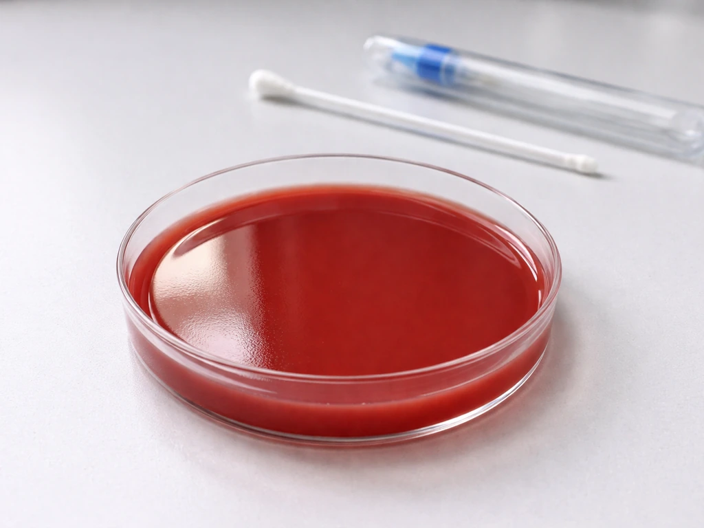
Blood agar is made by adding whole or defibrinated red blood cells (usually 5% sheep blood) to a nutrient agar base, typically tryptic soy agar (TSA) or Columbia agar base, after autoclaving and cooling it to about 48°C. The FDA's Bacteriological Analytical Manual, for example, describes mixing 5 mL of defibrinated sheep red blood cells into 100 mL of melted blood agar base. The agar base itself already contains casein and soy peptones, which supply nitrogen, amino acids, vitamins, and short peptides that most bacteria need to build proteins and replicate. The blood component then layers on top of that, adding hemin (also called factor X), additional proteins, lipids, and carbohydrates from the red blood cell membranes and cytoplasm.
The practical result is a rich, all-around supportive environment. Think of it like a well-stocked kitchen versus a vending machine: most organisms can find what they need here. That is exactly why blood agar is used as a baseline medium in clinical and teaching labs, not to isolate one type of organism, but to confirm that something can grow and to observe colony morphology and hemolysis patterns.
Gram-positive bacteria on blood agar
Gram-positive bacteria grow very well on blood agar, and this is where the medium really shines as a teaching tool. A blood agar plate is therefore a culture medium on which only Gram-positive organisms grow Gram-positive bacteria grow very well on blood agar. The thick peptidoglycan cell wall of Gram-positive organisms does not create any special nutritional requirement that blood agar fails to meet.
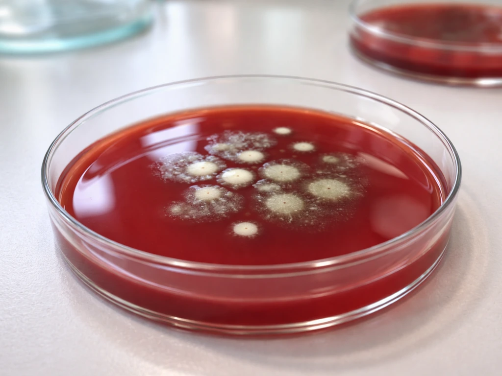
Classic examples include Staphylococcus aureus, which produces large, golden, beta-hemolytic colonies; Streptococcus pyogenes (Group A Strep), which produces small colonies with a clear zone of beta-hemolysis around them; and Enterococcus faecalis, which typically shows alpha-hemolysis or no hemolysis. Streptococcus grows on blood agar, where its nutritional needs and incubation conditions are well supported. Bacillus species, Listeria, and Clostridium (under anaerobic conditions) all grow on blood agar as well.
The hemolysis pattern is one of the most useful things blood agar reveals for Gram-positive cocci in particular. Beta-hemolysis means the organism has lysed the red blood cells completely, leaving a clear halo. Alpha-hemolysis produces a greenish, partial breakdown zone. No hemolysis (gamma) means the colonies just sit there without affecting the blood cells at all. These patterns help narrow down identity before you even do a Gram stain.
Gram-negative bacteria on blood agar
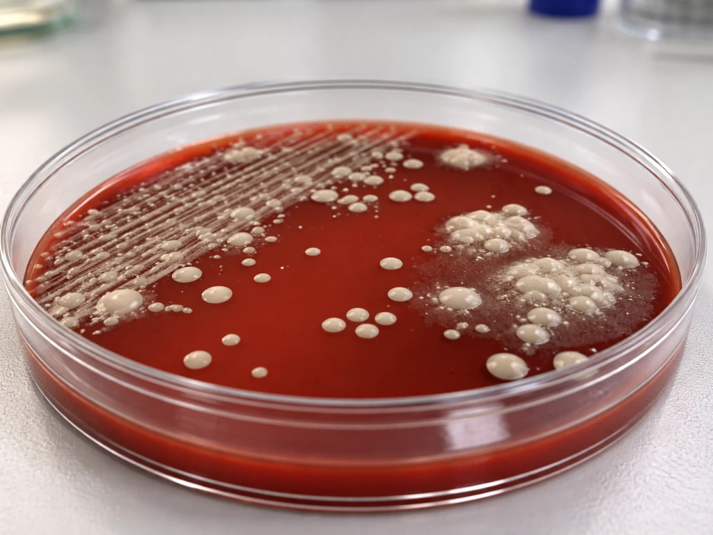
This is where a common misconception trips up students: many people assume blood agar is mainly for Gram-positive organisms because selective media like MacConkey or EMB agar specifically target Gram-negatives. That is backwards logic. Blood agar is not selective at all. Gram-negative bacteria grow readily on it. Escherichia coli, Klebsiella pneumoniae, Pseudomonas aeruginosa, Proteus mirabilis, and Enterobacter species are all well-documented growers on blood agar. Most Streptococcus species can grow on blood agar, making it a common medium for culturing and observing them. Pseudomonas, for instance, produces large flat colonies with a distinctive metallic sheen and can show beta-hemolysis. Proteus species famously swarm across the plate in concentric waves.
One important nuance: some fastidious Gram-negative organisms need more than what standard blood agar provides. Haemophilus influenzae, for example, requires both hemin (factor X) and NAD (factor V) to grow. Blood agar contains hemin from the red blood cells, but NAD is locked inside intact red blood cells and is not freely available on standard blood agar. That is why Haemophilus grows much better on chocolate agar, where the blood cells have been lysed by heat, releasing their contents.
Similarly, Neisseria gonorrhoeae prefers enriched selective media. So when a Gram-negative organism fails to grow on blood agar, the question to ask is whether the organism has unusual growth factor requirements, not whether blood agar is incompatible with Gram-negative bacteria as a class.
Growth depends on more than Gram reaction
The Gram stain tells you about cell wall structure. It does not tell you what nutrients an organism needs, what temperature it prefers, or whether it can tolerate oxygen. These are the factors that actually determine whether a given bacterium grows on any medium, including blood agar. This is a point that StatPearls (NCBI Bookshelf) makes explicitly: Gram stain reflects cell-wall properties and is a separate question from what a medium provides.
Here are the conditions that actually govern growth on blood agar:
- Temperature: Most lab protocols incubate blood agar plates at 35 to 37°C for 18 to 24 hours. Organisms with different optimal temperatures (like cold-loving psychrophiles or heat-adapted thermophiles) may grow poorly or not at all at standard lab temperatures, regardless of Gram reaction.
- Oxygen requirements: Standard blood agar plates incubated in ambient air favor aerobes and facultative anaerobes. Strict anaerobes like Clostridium require an anaerobic chamber or jar. Microaerophiles like Campylobacter need reduced oxygen with elevated CO2. The same plate, same medium, different atmosphere, very different results.
- Special growth factors: As noted with Haemophilus, some organisms need specific molecules (hemin, NAD, X and V factors) that blood agar may not supply in sufficient quantities. Chocolate agar is essentially blood agar taken one step further, with the cells lysed to release these factors.
- pH: Blood agar base is buffered to approximately neutral pH (around 7.0 to 7.4), which suits most clinical isolates. Acid-loving organisms like Lactobacillus prefer lower pH environments and may grow weakly.
- Moisture and plate freshness: Dried-out plates or plates stored improperly can inhibit growth mechanically, independent of what the medium chemically provides.
It is also worth noting that blood agar is non-selective, meaning it does not contain inhibitors like bile salts or crystal violet that would suppress certain organisms. Selective media like MacConkey agar are a different category entirely, designed to inhibit Gram-positive bacteria while letting Gram-negatives through. Blood agar makes no such distinction. This connects to a broader principle: most Gram-positive and Gram-negative organisms will grow on non-selective enriched media when conditions are right, and blood agar is one of the most permissive options available.
A quick comparison: who grows, who struggles, and why
| Organism | Gram reaction | Grows on blood agar? | Key reason |
|---|---|---|---|
| Staphylococcus aureus | Positive | Yes, readily | Nutritionally undemanding; produces beta-hemolysis |
| Streptococcus pyogenes | Positive | Yes, readily | Needs enriched medium; blood agar provides it |
| Enterococcus faecalis | Positive | Yes | Grows well; usually alpha or gamma hemolysis |
| Escherichia coli | Negative | Yes, readily | Facultative anaerobe; not fastidious |
| Pseudomonas aeruginosa | Negative | Yes | Aerobe; grows well, sometimes with beta-hemolysis |
| Klebsiella pneumoniae | Negative | Yes | Facultative anaerobe; robust grower |
| Haemophilus influenzae | Negative | Poorly or not at all | Needs free NAD (factor V); lysed blood in chocolate agar required |
| Neisseria gonorrhoeae | Negative | Poorly | Fastidious; prefers selective enriched media like Thayer-Martin |
| Clostridium perfringens | Positive | Yes, under anaerobic conditions | Strict anaerobe; needs anaerobic incubation |
| Campylobacter jejuni | Negative | Poorly at standard conditions | Microaerophile; needs reduced O2 and elevated CO2 |
What to check when you see growth (or no growth)
When you plate something on blood agar and get results, here is a practical framework for interpreting what you see. Think of it as a short checklist that moves from the medium itself outward to the organism's biology.
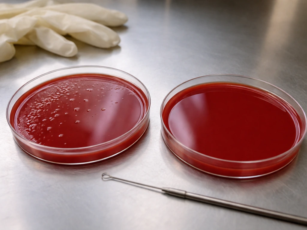
- Did you get any growth at all? If yes, the organism's basic nutritional needs are met by the peptones, amino acids, and blood components. If no, ask whether conditions (temperature, atmosphere, incubation time) were correct before concluding the organism cannot grow here.
- What does the hemolysis look like? Beta-hemolysis (clear halo) is the most dramatic and narrows your list significantly. Alpha-hemolysis (green tinge) suggests partial lysis. No hemolysis (gamma) is common in many Gram-negatives and some Gram-positives. Hemolysis pattern is a biological clue, not a Gram-reaction proxy.
- What is the colony morphology? Size, color, texture, and odor can all point toward specific genera before you do any further testing. Pseudomonas smells distinctly fruity. Proteus spreads in waves. Staphylococcus produces pigmented, raised, round colonies.
- Now do the Gram stain. The stain tells you cell-wall structure (positive = thick peptidoglycan, negative = thin peptidoglycan with outer membrane) and cell shape, which combined with hemolysis pattern and colony morphology puts you in a very specific part of the bacterial world.
- If growth was poor or absent, consider the organism's special requirements. Does it need factors not available in standard blood agar, like free NAD for Haemophilus? Does it need anaerobic conditions? Is the incubation temperature wrong? Growth failure on blood agar is usually a conditions problem, not a fundamental incompatibility.
One more thing worth keeping in mind: blood agar is often used alongside selective media in real workflows precisely because it shows you everything that can grow, while the selective plate shows you the subset you are hunting for.
If you are trying to identify an unknown organism for a class exercise or lab project, starting with blood agar gives you the full picture of what is present, and then you use the Gram stain and other biochemical tests to narrow it down.
A helpful way to think about this is the broader question of why bacteria grow on agar in the first place: they grow when the medium supplies the nutrients and conditions they need why does bacteria grow on agar. The medium is your first step, not your last.
In the same way, you can also ask which bacterial mixtures can grow as a biofilm under the conditions you are testing which of the bacterial mixtures can grow as a biofilm.
The bottom line on blood agar and bacterial growth
Blood agar does not pick sides in the Gram-positive versus Gram-negative debate. It supports both, because its enriched formulation of peptones, amino acids, and blood components meets the fundamental nutritional needs of the vast majority of bacteria you will encounter in a clinical or educational context. The organisms that struggle on blood agar are usually fastidious in specific ways: they need growth factors that become available only when red blood cells are lysed (like Haemophilus), or they need unusual atmospheric conditions (like strict anaerobes or microaerophiles), or they have temperature preferences that differ from standard incubation. Understanding those exceptions is actually the deeper lesson here: growth on any medium is a product of the intersection between what the medium provides and what the organism biologically requires, and Gram reaction is only one small piece of that picture.
FAQ
If a bacterium is Gram-negative, will it always grow on blood agar?
Yes, some organisms grow poorly or not at all on standard blood agar even if they are otherwise “nonselective.” Common reasons include strict anaerobes needing an anaerobic atmosphere, microaerophiles needing reduced oxygen, or temperature preferences that differ from typical 35 to 37°C. If you suspect one of these, incubating under the correct atmosphere or temperature (or using a specialized medium) is usually the next step.
Why might I not see clear hemolysis even when an organism typically shows beta-hemolysis on blood agar?
Not necessarily. Blood agar is generally enriched, but the hemolysis pattern depends on the organism and its virulence factors, and the appearance can be affected by incubation time and plate age. A weak or delayed hemolytic reaction can look like partial hemolysis if the plate is read too early, or it can diminish if the agar dried out or if the blood component degraded.
What should I consider if my suspected Haemophilus or other fastidious bacteria fail to grow on blood agar?
If you are working with fastidious organisms, standard blood agar may lack growth factors that are only released when red cells are lysed. For example, Haemophilus influenzae often grows better on chocolate agar because heating the blood releases factors that are not freely available on intact-cell blood agar. So failure to grow on blood agar should prompt consideration of factor requirements, not a conclusion about Gram type.
Can strict anaerobes grow on blood agar, and how does incubation affect what I see?
Yes, but it will generally be slower or more variable, and colony morphology can be harder to compare. Growth on blood agar depends on atmosphere, and many organisms tolerate oxygen only partially. If you need to support anaerobes, using an anaerobic jar or chamber and reading plates at appropriate time points helps avoid false negatives.
How reliable is colony shape and hemolysis on blood agar for identifying an unknown organism?
Colony morphology and hemolysis are helpful, but there are look-alike situations. For instance, different genera can produce colonies that look similar in size and shape, and some organisms can show atypical hemolysis. The safest approach is to combine blood agar findings with a Gram stain and targeted biochemical or molecular tests rather than relying on hemolysis alone for identification.
Do different blood agar brands or blood sources change growth or hemolysis results?
The volume and type of blood and the agar base formulation matter for how robust the growth is and how hemolysis appears. Plates made with fresh versus older blood, different animal blood sources, or different agar bases can change clarity and the intensity of hemolytic halos. If results are inconsistent, verifying the plate batch or using a standardized commercial plate can improve comparability.
What problems can happen if my blood agar plate has a mixed bacterial sample?
Mixed cultures can obscure interpretation. Overgrowth by one fast-growing organism may suppress slower growers, and hemolysis can look smeared or generalized across the plate. If you suspect a mixed sample, repeating with appropriate dilution streaking, using selective media alongside blood agar, and confirming with microscopy can reduce misreads.
How does incubation time and plate reading schedule affect hemolysis interpretation on blood agar?
Hemolysis requires that the organism actually reach enough viable density at the time of reading and that the reading is timed correctly. If you incubate too short, weak hemolysis may be missed; if you wait too long, cells may lyse and change the background appearance. Following standard incubation and using consistent time points for reading helps.