Bacteria grow on agar plates because agar gives them exactly what they need to settle, replicate, and form visible colonies: a stable, moist solid surface to anchor to, and (when the agar is nutrient-supplemented) the carbon, nitrogen, and minerals required for metabolism. Plain agar on its own is just a gelling agent with no nutritional value, so bacteria can only thrive on it if they either brought their own nutrients along or the agar has been formulated with a nutrient base like tryptic soy broth, blood, or MacConkey salts. For Staphylococcus aureus, the commonly used choices are nutrient-rich media such as blood agar or other formulated agar plates. Get the surface, the nutrients, and the incubation conditions right and you'll see colonies. Get any one of those wrong and the plate stays blank.
Why Does Bacteria Grow on Agar Plates and How to Fix It

What agar actually is and why it makes a good home for bacteria
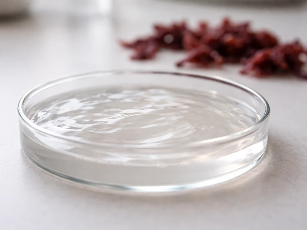
Agar is a polysaccharide derived from red algae. Microbiologists use it almost universally as a solidifying agent because bacteria cannot digest or degrade it, so the surface stays intact throughout incubation. That matters a lot: if bacteria could eat the scaffold, the whole plate would liquefy and you'd lose your colony structure. Agar also melts at around 85°C and solidifies around 40°C, which means you can pour it hot, let it set, and then inoculate it at room temperature without harming the cells.
The physical structure of a set agar plate creates what researchers describe as a solid/air-gel interface. That interface does something important: it supports bacterial adhesion. Cells landing on the surface can attach, resist being washed away, and begin dividing in place. The gel matrix also holds moisture, which bacteria absolutely require to carry out any metabolic activity. Without that moisture reservoir, cells would desiccate and die before they ever formed a colony. So even before you count a single nutrient molecule, the physical properties of agar are already doing critical work.
From inoculation to visible colony: what actually happens on the plate
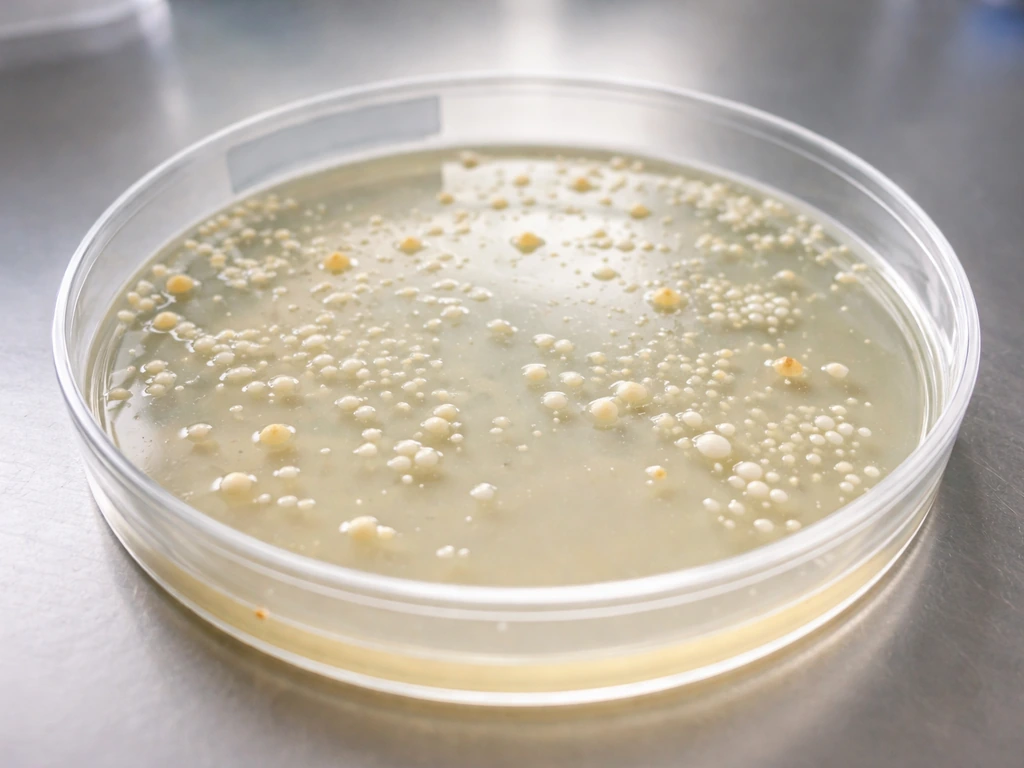
When you streak a loop or spread a diluted sample across an agar plate, you're depositing individual bacterial cells onto that gel surface. Each viable cell that lands in an isolated spot becomes the founder of a colony. Over the next 24 to 48 hours of incubation, that single cell divides repeatedly, and because the cells stay anchored on the solid surface rather than dispersing into liquid, all the daughter cells pile up in one spot. The result is a visible mound of genetically identical cells called a colony. Some bacterial mixtures also show different behavior in biofilm-forming conditions, where cells adhere and grow in structured communities rather than as single dispersed colonies.
Two practical variables control whether those founders actually produce visible colonies: inoculum size and incubation time. Research tracking soil bacteria on solid plates found that both factors significantly affected culturability, with some colonies only becoming visible after weeks of extended incubation. For most routine lab bacteria like E. coli or Staphylococcus aureus, 24 to 48 hours at the right temperature is plenty. But for slow-growing or nutrient-stressed organisms, you may need to wait much longer before concluding a plate is negative. The colony you see is not just "bacteria growing on agar" in a vague sense. It's a three-dimensional structure shaped by nutrient gradients, oxygen diffusion from the surface, and the rate at which the founding cell was able to replicate.
Nutrient agar vs. plain agar: not all plates are equal
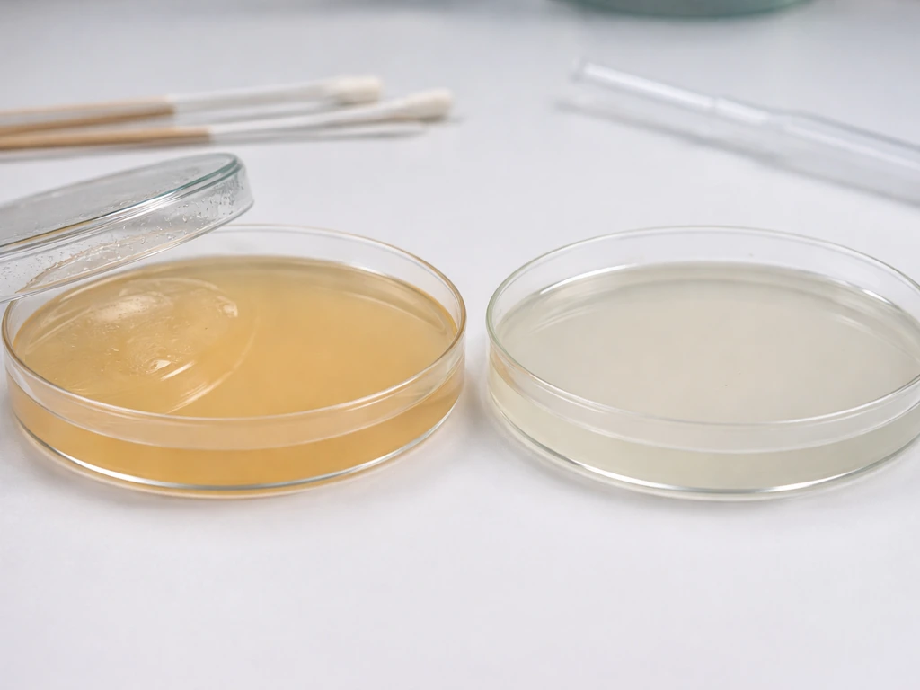
This is probably the most common source of confusion for students and beginners. "Agar" by itself is not food for bacteria. If you dissolved agar in distilled water, poured plates, and inoculated them, most bacteria would struggle or fail to grow because there's nothing to metabolize. The agar is just the scaffold. The nutrients have to be built into the formulation. Selective media can be formulated so that only gram-positive organisms grow, which helps identify them while suppressing gram-negative bacteria nutrients have to be built into the formulation.
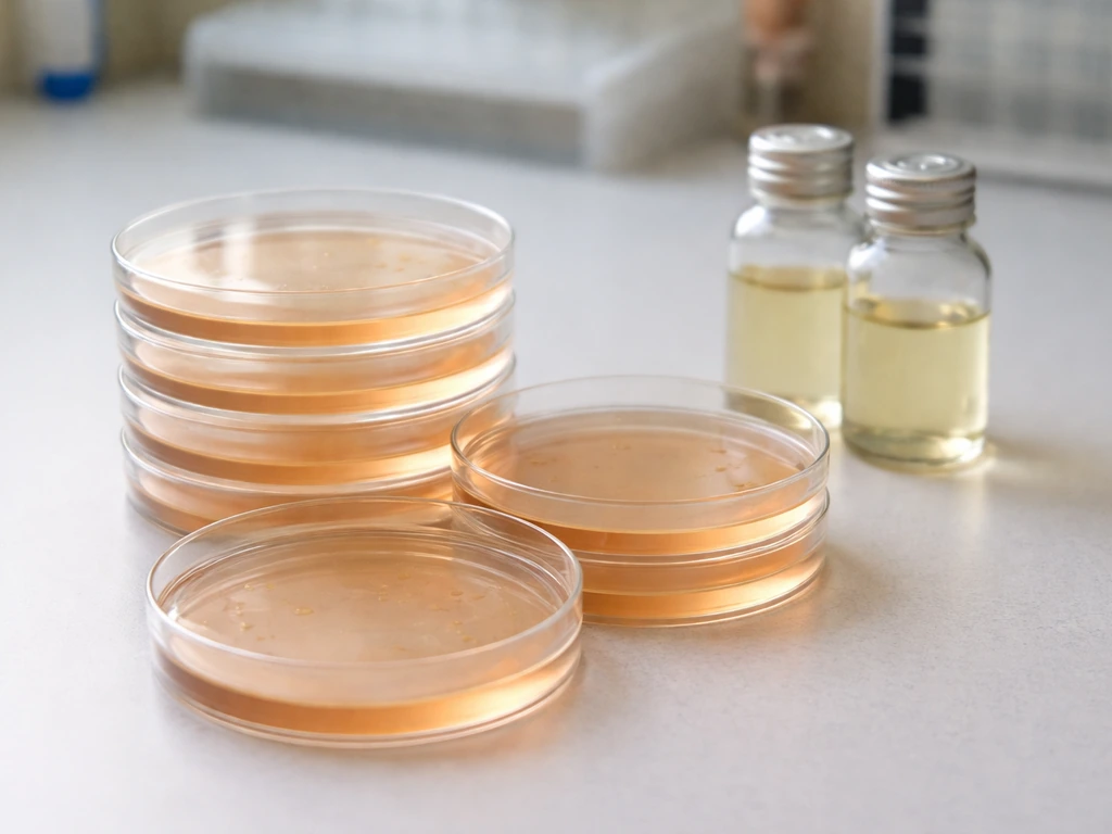
Most standard lab plates are nutrient agar or tryptic soy agar (TSA), which contain peptones, beef extract, or casein digests that supply carbon and nitrogen. Blood agar adds red blood cells to support even fastidious organisms that need specific growth factors. Selective agars like MacConkey go further by adding inhibitors that block certain bacteria while allowing others to thrive. Understanding which agar type you're using is the first diagnostic question to ask if your plate isn't behaving the way you expect. The question of which organisms grow on which formulation comes up constantly in practice: blood agar supports streptococci and many fastidious pathogens that won't grow on plain nutrient agar, while selective and differential agars are designed to filter the microbial population down to specific groups.
| Agar Type | Key Nutrients/Additives | Who Grows | Best Used For |
|---|---|---|---|
| Plain/simple agar | None (agar + water only) | Almost nothing | Demonstrating why nutrients matter |
| Nutrient agar | Peptone, beef extract | Most non-fastidious bacteria | General cultivation |
| Tryptic soy agar (TSA) | Casein and soy peptones | Wide range of bacteria and some fungi | Broad-spectrum isolation |
| Blood agar | Nutrient base + 5% sheep blood | Fastidious organisms, streptococci | Clinical and fastidious culture |
| MacConkey agar | Bile salts, crystal violet, lactose | Gram-negative bacteria only | Selective and differential work |
| Mannitol salt agar | High salt, mannitol, phenol red | Salt-tolerant organisms (e.g., Staph. aureus) | Selective isolation from mixed samples |
The conditions that actually control whether bacteria grow
Nutrient agar and a sterile technique get you partway there, but four environmental conditions determine whether bacteria actually multiply on that plate. Think of these as dials you need to have set correctly simultaneously. Getting three out of four right still might mean no growth.
Temperature
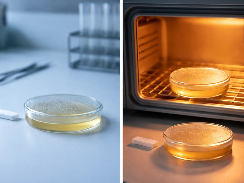
Most human-associated pathogens are mesophiles, meaning they grow optimally between 35°C and 37°C, which is close to body temperature. Leave those plates at room temperature (around 20-22°C) and growth will be dramatically slower or may not appear within your expected timeframe. Incubate at 4°C (a refrigerator) and growth halts almost entirely for most species. On the other end, incubating above 45°C will denature the enzymes of most mesophiles and kill the cells. Colony radial growth rate studies confirm that temperature is one of the primary variables controlling how fast colonies expand on solid medium. This is also why your incubator temperature needs to be verified regularly, not just assumed.
pH
Most bacteria prefer a pH close to neutral, typically between 6.5 and 7.5. Standard nutrient and TSA plates are buffered to sit right in that range. If your agar was prepared incorrectly, contaminated with something acidic, or stored in conditions that shifted its pH, you may see poor or patchy growth. Some organisms, like Lactobacillus species, tolerate lower pH, but for the vast majority of common lab bacteria, stray significantly from neutral and the plate will underperform.
Moisture and water activity
Bacteria need free water for every biochemical reaction inside the cell. Agar plates that have been left to dry out (especially plates stored for too long or incorrectly) have reduced water activity, and growth will be sparse or absent. Humidity in the gas phase surrounding the plate also matters: colony growth rate studies show that the humidity of the environment around the plate directly affects how quickly colonies expand. This is why incubators often have water trays to maintain humidity, and why you should store poured plates sealed and refrigerated rather than loose on a bench.
Oxygen requirements
Agar plates exposed to open air are aerobic environments by default, which is fine for obligate aerobes and facultative anaerobes (organisms that can use oxygen but don't require it). Because Streptococcus species are typically grown on nutrient media such as blood agar (or other enriched agars) rather than plain agar, the plate type matters as much as the environmental conditions. Obligate anaerobes, however, will not grow on open plates because oxygen is toxic to them. If you're trying to culture anaerobic bacteria like Clostridium species, you need an anaerobic chamber or jar that scrubs oxygen from the environment. Conversely, oxygen diffusion from the plate surface into the agar affects how deep colonies can develop in the gel, which connects back to the three-dimensional structure of colony formation.
Troubleshooting your agar plates when growth looks wrong
Whether you're getting no growth when you expect it, or unexpected growth when you don't, the checklist below covers the most common culprits. Work through these systematically rather than changing multiple variables at once.
No growth or very sparse growth
- Wrong agar type: check whether your plate actually contains nutrients, or whether you accidentally used plain water agar or a selective agar that inhibits your target organism
- Incubation temperature off: verify your incubator with a calibrated thermometer, not just the dial reading
- Dried-out plates: plates stored too long or at low humidity lose water activity; use fresh plates within a few weeks of pouring
- Inoculum too small or not viable: if you inoculated from an old culture, broth that was improperly stored, or a sample that was exposed to heat or disinfectant, the cells may be dead
- Incubation time too short: slow-growing organisms may need 48-72 hours or more; some environmental bacteria can take weeks to form visible colonies
- Anaerobic organism on an aerobic plate: if you're working with known anaerobes, standard plate incubation won't work
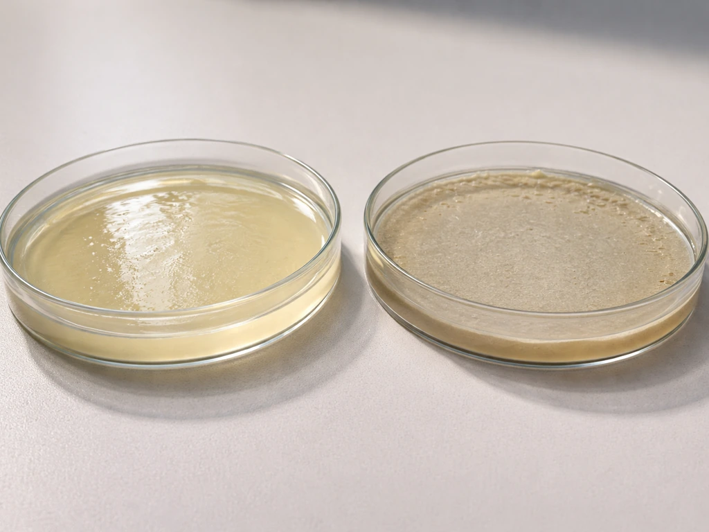
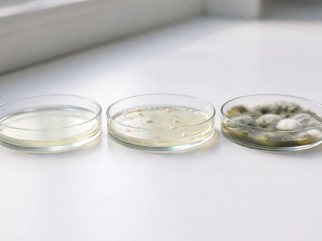
Unexpected or unwanted growth (contamination)
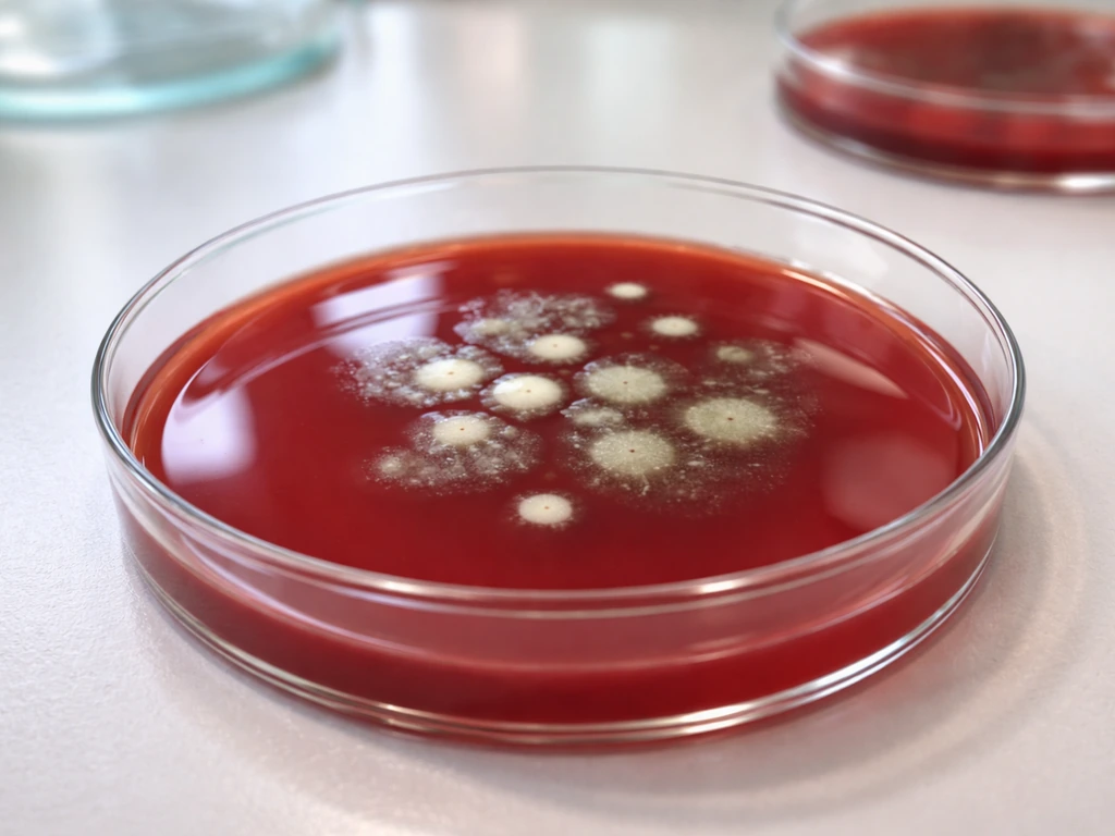
- Fuzzy or spreading growth with a different texture than bacterial colonies usually indicates fungal contamination from airborne mold spores
- Growth in uninoculated areas of the plate suggests contamination during pouring, during inoculation, or from a non-sterile loop or swab
- Multiple colony types (different sizes, colors, morphologies) on a plate that should show a pure culture points to contamination or a mixed inoculum
- Growth on control plates (blank, uninoculated plates) that were incubated alongside your test plates is a clear sign of contamination in the agar itself or the pouring environment
Aseptic technique: how to keep contamination off your plates
Aseptic technique is the set of practices that prevent unwanted microorganisms from getting onto your agar plates and ruining your results. The basic principle is simple: everything that touches your sterile agar or your inoculum needs to be sterile itself, and you need to minimize exposure to the non-sterile air and surfaces around you.
- Work near a flame or inside a laminar flow hood: heat from a Bunsen burner creates an upward convection current that keeps airborne particles from settling onto your open plates
- Sterilize your inoculating loop by flaming it to red-hot before and after use, or use disposable sterile loops
- Keep plate lids on as much as possible: only open the plate when you are actively inoculating, and angle the lid to act as a shield rather than setting it flat on the bench
- Pour plates in a clean environment: if you're preparing agar from scratch, pour it shortly after autoclaving while the agar is still liquid (around 50°C), and do so quickly
- Label plates on the bottom, not the lid: lids can be swapped accidentally, leading to misidentified results
- Incubate plates upside down (agar side up): this prevents condensation from dripping onto the colony surface and spreading or distorting colonies
Even with good technique, airborne contamination happens sometimes, especially in non-laboratory settings like classrooms. Running a negative control (an uninoculated plate incubated under identical conditions) lets you distinguish between real growth from your inoculum and background contamination from the environment. If your negative control shows growth, your results from that batch of plates are unreliable regardless of what else you see.
Putting it all together: why agar works and how to make it work for you
Bacteria grow on agar because the gel surface gives them a place to anchor, the moisture keeps their chemistry running, and (when nutrients are present) the plate feeds their metabolism well enough for repeated cell division. A single viable cell can multiply into a visible colony of millions in 24 to 48 hours under good conditions. That's the core mechanism. Everything else, temperature, pH, oxygen, inoculum size, agar formulation, is a dial that either enables or prevents that process from happening.
If you're diagnosing a plate problem today, start with the agar type (does it actually contain nutrients for your organism?), then verify your incubation temperature, then assess the age and hydration of your plates, and finally scrutinize your technique for contamination opportunities. Most plate failures come down to one of those four things. And if you see unexpected colony types, sizes, or distribution patterns, that's usually a contamination signal, not a biological mystery. Understanding these fundamentals makes it much easier to interpret results from more specialized media too, whether you're looking at what grows on blood agar, what selective agars block, or how different gram-positive and gram-negative organisms respond to different formulations.
FAQ
If bacteria need nutrients, why do colonies appear on a plate that is labeled only “agar”?
Most agar plates fail because they are either nutrient-free (plain gelling agent) or because the plate’s nutrient formulation does not match the organism’s needs. If you are using a “general purpose” plate, confirm it is nutrient agar or TSA, then check whether your target organism is fastidious (may require enriched media like blood agar).
How do I tell whether colonies are from my sample or just contamination?
A negative control is inoculum-free but incubated the same way, same incubator, same time window, and the same handling steps. If that control shows colonies, treat your result as contamination and do not interpret those colonies as coming from your sample.
Can an agar plate that seemed blank become positive after more incubation time?
Yes. If you incubate too long, stressed or slow growers can eventually appear, making a “negative” plate look positive later. Keep your read time consistent with your organism expectations (for many common lab bacteria this is often within 24 to 48 hours), then record any later appearances as separate observations.
If my streak came out blank, does that always mean my sample had no bacteria?
A small number of cells can produce a visible colony even if your inoculum is low, but “no growth” can still happen if the plate is dried, too cold, has an off pH, or lacks the right nutrients. To distinguish inoculum size from plate conditions, compare against a known positive control organism on the same batch of plates.
Why do I get a “lawn” instead of isolated colonies when I streak on agar?
Colony morphology patterns, such as one spot with dense growth versus evenly spread colonies, can indicate whether cells were properly isolated. Overloading the sample or not mixing during dilution often leads to confluent growth (a lawn) rather than discrete colonies, which can mask differences between species.
Why won’t obligate anaerobes grow on standard open agar plates?
For facultative anaerobes and aerobes, colonies can appear on the surface of an open plate because oxygen is available. For obligate anaerobes, oxygen exposure prevents growth, so you need an oxygen-free setup (anaerobic jar or chamber) and appropriate anaerobic media.
What’s the real impact of incubating at the wrong temperature, not just “faster” or “slower” growth?
Wrong temperature can change both growth speed and colony appearance. Even if growth happens, incubating outside the organism’s expected range can select for atypical survivors, shift colony sizes, or alter metabolic states, which affects interpretation.
How do I choose the correct agar type when a plate is not working?
Using the wrong plate type can cause apparent “no growth” even when bacteria are present. Confirm the medium is not only nutrient-rich but also compatible with the organism, and that selective or differential additives (inhibitors, salts, blood components) are intended for your target.
How can plate storage and age cause poor growth even if the medium type is correct?
Yes, plate handling matters. Repeated warming, improper storage, or plates that are too old can lose moisture or have altered water activity, and this often produces smaller, sparse, or delayed colonies compared with fresh, properly sealed plates.
What practical mistakes during pouring or incubation setup can lead to patchy or weird colony patterns?
If your plates were poured or handled at the wrong step, you can create problems like uneven solidification, surface cracks, or excessive dryness. Uneven surfaces can also trap cells inconsistently, leading to patchy growth that looks like “biology” rather than a technical issue.