Yes, bacteria can grow on potato dextrose agar (PDA), even though PDA is specifically designed to cultivate fungi. The medium's nutrient-rich composition and mildly acidic pH of around 5.6 do not reliably stop many environmental, soil, or plant-associated bacteria. The result: if your technique is not tight, or if your sample comes from a microbe-rich environment, bacterial colonies will appear on your PDA plates alongside, or even instead of, the fungi you were trying to grow.
Can Bacteria Grow on PDA? Reasons, Recognition, Prevention

Marcus Holloway
20 Jul 2026
What is potato dextrose agar and why is it formulated for fungi?
PDA is one of the most widely used mycological media in teaching labs and research settings worldwide. A standard from-scratch recipe calls for a filtrate made by boiling 300 g of diced potatoes in 500 mL of water, then bringing that filtrate up to 1 liter, adding 20 g of glucose (dextrose) and 15 g of agar per liter. Commercially, products like BD Difco PDA come as a dehydrated powder at 39.0 g per liter, and after autoclaving at 121°C the final pH settles at around 5.6 (±0.2 at 25°C, per Sigma-Aldrich specifications). The potato infusion provides a complex mix of amino acids, vitamins, and trace minerals, while the dextrose is a quick-access carbon and energy source. Agar is the solidifying agent and has no nutritional role.
The reason mycologists reach for PDA first is that fungi genuinely thrive in mildly acidic, carbohydrate-rich environments. Most bacteria, by contrast, grow best at neutral to slightly alkaline pH (near 7.0). So a pH of 5.6 gives fungi a competitive edge, and the complex potato nutrients support the slower, more sprawling growth typical of filamentous molds and yeasts. PDA is particularly valued for inducing sporulation and showing characteristic colony morphology, which is essential when you need to identify a fungal species by its reproductive structures. In short, PDA is selective not because it kills bacteria outright, but because it tilts chemical conditions in favor of fungi.
Why bacteria sometimes grow on PDA anyway
Think of PDA's pH 5.6 as a gentle preference, not a locked door. Several overlapping factors determine whether bacteria will establish themselves on a PDA plate.
Nutrients
The potato infusion and 20 g/L of glucose make PDA genuinely nutrient-rich. Many environmental bacteria are metabolically flexible enough to exploit those nutrients at pH 5.6, especially if they are already adapted to slightly acidic soils or plant surfaces. A high glucose concentration in particular is a ready carbon source for fast-growing bacteria like Pseudomonas species, which have been recovered on PDA in comparative cultivation studies.
pH
A pH of 5.6 is mildly acidic, but it is not strongly bacteriostatic. Many Gram-negative rods and spore-forming Gram-positive bacteria tolerate this range without difficulty. The real bacterial suppression only kicks in when PDA is further acidified, dropping the pH to around 3.5 with sterile tartaric or citric acid. That level of acidification is a recognized mycology technique for isolating fungi from heavily contaminated environmental samples, but standard commercial PDA does not go anywhere near that range.
Temperature
Standard mycology protocols incubate PDA plates at 20 to 25°C to favor fungal sporulation and colony development. Many clinical or enteric bacteria prefer 30 to 37°C, so cooler incubation does reduce bacterial competition. However, psychrotolerant bacteria such as Pseudomonas species or Listeria monocytogenes can grow efficiently at 20 to 25°C, and even at refrigerator temperatures around 4°C. So cooler incubation helps but is not a reliable sole barrier.
Oxygen and moisture
Standard PDA plates are incubated aerobically, which supports aerobes and facultative anaerobes, the two groups that dominate most environmental and clinical bacterial populations. BMBL (CDC/NIH), principles on oxygen requirement and anaerobic culture note that obligate anaerobes require anaerobic media and an oxygen‑free environment to grow and normally will not be recovered from PDA incubated aerobically blank" rel="noopener noreferrer">BMBL (CDC/NIH) — principles on oxygen requirement and anaerobic culture. Obligate anaerobes cannot grow under these conditions, but they are a minority in the settings where PDA is typically used. Moisture matters too: PDA solidified with 15 g/L agar has a high water activity, which bacteria need to proliferate. This is a key difference from surfaces like glass or silicone, where water availability depends entirely on the environment around them.
Inoculum source
Perhaps the most underappreciated factor is where the sample came from. A soil core, a decaying plant, a water sample, or an unwashed hand carries a massive and diverse bacterial load. Even with perfect technique, environmental samples can transfer thousands of bacterial cells onto a plate for every fungal spore. Under those conditions, the competitive and numerical advantage of bacteria means that bacterial colonies can dominate a PDA plate before your target fungus has had time to establish.
Which bacteria can grow on PDA and which usually cannot
Not all bacteria are equally likely to appear as contaminants on PDA. Their success depends on acid tolerance, nutritional flexibility, and temperature preference. Here is a practical breakdown.
| Bacterial Group | Can It Grow on Standard PDA? | Key Reason |
|---|---|---|
| Pseudomonas spp. (environmental/plant) | Yes, commonly | Acid-tolerant, metabolically versatile, grows at 20–25°C |
| Bacillus spp. (soil spore-formers) | Yes, frequently | Broad pH and temperature tolerance; heat-resistant endospores survive autoclaving if medium is re-contaminated |
| Streptomyces and actinobacteria (soil) | Yes, on modified or standard PDA | 2024 studies show PDA can support actinobacterial cultivation with formula adjustments |
| Staphylococcus spp. | Sometimes | Tolerates mild acidity; grows aerobically but prefers neutral pH and 37°C |
| Listeria monocytogenes | Possible at cooler incubation temps | Psychrotrophic; can grow near 4°C and at 20–25°C |
| Enteric bacteria (E. coli, Salmonella) | Less common; possible with heavy inoculum | Prefer neutral pH and 37°C; less competitive at 20–25°C and pH 5.6 |
| Strict anaerobes (Clostridium, Bacteroides) | No under standard aerobic conditions | Oxygen-sensitive; standard PDA incubation is aerobic |
| Highly acidophilic species (Acidithiobacillus) | Theoretically yes, but rarely relevant in lab contamination | pH 5.6 is not restrictive enough to exclude them, but they are not typical lab contaminants |
The practical takeaway is that soil and plant-associated genera like Pseudomonas and Bacillus are your most common unwanted guests on a PDA plate in a teaching lab. Fast-growing Pseudomonas colonies in particular can spread across a plate quickly and physically crowd out slower fungal growth, so early detection matters.
What colonies look like and how to tell bacteria from fungi
One of the most useful skills in a microbiology lab, whether you are a student or a seasoned researcher, is being able to look at a plate and make a reasonable first call about what you are looking at. Visual inspection is never definitive, but it guides your next step.
Visual cues at a glance
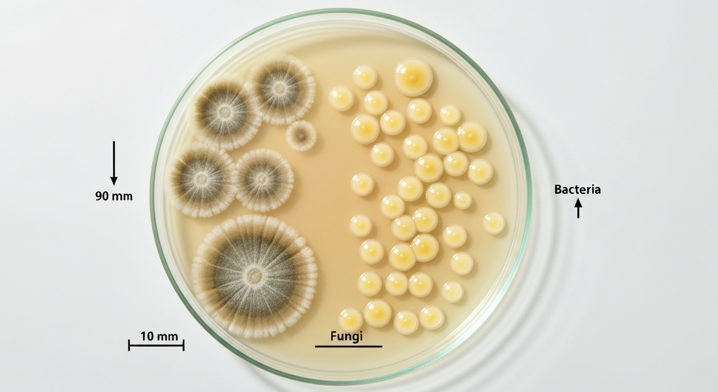
- Fungal colonies: typically show radial, filamentous, or fuzzy mycelial growth; surface texture may be powdery, woolly, or velvety as spores form; colony color often changes over time and may show distinct pigmentation on the front and reverse of the plate; growth tends to be slower, spreading outward in a circular pattern.
- Bacterial colonies: usually appear smooth, moist, or creamy with a glossy surface; some (like Bacillus) can be dry, wrinkled, or flat with irregular edges; shape tends to be compact and convex rather than spreading; growth appears faster, sometimes within 24 to 48 hours at 20–25°C.
- Yeast colonies (fungi but non-filamentous): can resemble bacteria in that they are smooth and moist, but they are generally larger, creamier, and sometimes slightly mucoid; this is a common source of confusion for newer students.
Confirmatory checks you can do in a teaching lab
Visual inspection alone is not enough to make a final call. The first confirmatory step is a simple wet mount. For a suspected fungal colony, a drop of lactophenol cotton blue (LPCB) stain on a coverslip preparation will reveal hyphal structures, conidia, and other reproductive features that are unmistakably fungal. For a suspected bacterial colony, a Gram stain is the standard workhorse: it reveals cell shape (cocci, rods, filaments), arrangement (clusters, chains, pairs), and Gram reaction (positive or negative), none of which fungi will show in the same way. A KOH or calcofluor white mount is another quick option for highlighting fungal cell walls.
If you are still uncertain, subculture the suspect colony. Transfer it to a general-purpose bacterial medium like tryptic soy agar (TSA) or nutrient agar and incubate at 30 to 37°C: if it grows well under those conditions and produces typical bacterial colony forms, you have confirmed bacterial contamination. ATCC Mycology Culture Guide (handling and subculture recommendations) recommends transferring suspected bacterial colonies to bacterial media (e.g., nutrient agar or TSA) and incubating at bacteriology temperatures (30–37°C) to confirm bacterial growth characteristics. Conversely, subculturing onto fresh PDA and performing an LPCB mount on the result helps confirm a fungal identity. This cross-check approach is standard practice in both clinical mycology and teaching labs.
Practical ways to reduce bacterial contamination on PDA
Understanding why bacteria grow on PDA makes it much easier to choose the right countermeasure. Each intervention targets one or more of the factors described above.
Acidification
Lowering the pH of PDA from its standard 5.6 down to approximately 3.5 using a sterile solution of tartaric acid or citric acid is one of the oldest and most reliable methods for suppressing bacterial contaminants when isolating fungi from soil or plant material. At pH 3.5, most bacteria cannot maintain normal cellular function, while acid-tolerant fungi continue to grow. The key caution: add acid to cooled, molten PDA just before pouring plates (not before autoclaving, as autoclaving at low pH can hydrolyze the agar and destroy nutrients). Also note that this level of acidification can slow or inhibit some acid-sensitive fungi, so it works best when you already expect heavy bacterial contamination and are willing to accept some trade-off in fungal recovery.
Adding antibiotics
Commercial selective mycology media like Mycosel use a combination of chloramphenicol (approximately 50 µg/mL) to suppress a broad range of bacteria and cycloheximide (approximately 500 µg/mL) to suppress fast-growing environmental molds that might otherwise outcompete slower-growing fungi of interest, such as dimorphic pathogens. Chloramphenicol alone is commonly added to standard PDA when broad bacterial suppression is needed without restricting environmental fungi. These antibiotic concentrations are standard in clinical and environmental mycology. A practical warning for classroom settings: cycloheximide is toxic to mammals at low doses, so it requires careful handling and is generally inappropriate for unsupervised student use; chloramphenicol carries its own risks and should be handled with appropriate PPE and institutional guidance.
Incubation conditions
Incubating PDA plates at 20 to 25°C rather than 30 to 37°C disadvantages most mesophilic bacteria that thrive at warmer temperatures. This alone will not eliminate contamination from psychrotolerant species, but it meaningfully reduces the competitive window for the most common lab contaminants. Incubation duration also matters: checking plates at 48-hour intervals and removing or documenting contaminated plates before bacterial colonies overgrow helps preserve interpretable results.
Sterile technique
This is the foundation everything else rests on. Proper aseptic technique includes working near a flame or inside a biosafety cabinet, using sterilized loops and spatulas, not talking or coughing over open plates, and keeping plates covered whenever possible. Bacterial contamination on PDA is very frequently traced back to technique failures rather than medium failures. Reinforce this in every lab session, because even experienced scientists occasionally have contaminated plates from a single lapse in attention.
Controls and sampling strategies
Every experiment should include uninoculated PDA control plates exposed to the same environment (a settle plate left open during sample processing, for example) and sterility control plates kept sealed throughout. These tell you how much environmental contamination was introduced during the procedure versus how much came from the sample itself. Parallel plating on both standard PDA and acidified or antibiotic-supplemented PDA from the same sample allows direct comparison of bacterial versus fungal recovery, which is genuinely useful data, not just a redundancy.
How this compares to other bacterial growth environments
Bacterial growth on PDA is really a story about nutrients, pH, and available water working together. It helps to put PDA in context by comparing it to other environments that come up in microbiology education.
Distilled water is essentially the opposite of PDA in terms of nutrients. It provides essentially no carbon, nitrogen, or energy sources, and bacteria that persist in distilled water are surviving in a dormant or near-dormant state rather than actively growing the way they can on nutrient-rich PDA. If you ask whether distilled water will grow bacteria (does distilled water grow bacteria), the short answer is no for active growth, distilled water lacks the nutrients bacteria need, though some can survive in a dormant state. The comparison is useful because it illustrates that nutrient availability, not just pH or temperature, is a primary driver of active bacterial proliferation.
Refrigerator storage (around 4°C) is a concept that connects directly to the temperature discussion above. A PDA plate left in a refrigerator does not become sterile. Psychrotrophic bacteria like Pseudomonas and Listeria monocytogenes can still grow, albeit slowly, and PDA's nutrients remain fully available. For more detail on psychrotrophic organisms and refrigerator growth, see can bacteria grow in a fridge. This is why refrigerating plates is used to slow contamination during storage, not to prevent it entirely.
Silicone surfaces and glass surfaces are inert materials that provide no nutrients to bacteria on their own. Bacteria can adhere to both and potentially form biofilms, but active multiplication depends on nutrients arriving from elsewhere, such as organic residues or fluids in contact with the surface. For more detail on whether bacteria can grow on silicone, see can bacteria grow on silicone. PDA is fundamentally different because the medium itself is the nutrient source, making it a much more hospitable environment for growth than a clean glass or silicone surface.
A vacuum environment eliminates oxygen entirely. Standard PDA plates are incubated in ambient air, so obligate anaerobes cannot grow on them, as discussed earlier. In a true vacuum, even aerobic bacteria cannot grow because they require oxygen for respiration. PDA in a vacuum would be a hostile environment for most bacteria, which is essentially the inverse of what makes open, aerobically incubated PDA so permissive.
Safety and classroom protocols when working with PDA
Working with PDA in a classroom or teaching lab context is generally low-risk, but it comes with responsibilities that students and educators should take seriously.
PPE and handling
- Wear gloves and a lab coat when preparing, inoculating, or handling plates. Nitrile gloves are appropriate for standard PDA work without antibiotics.
- If using antibiotic-supplemented media, especially cycloheximide, wear additional protection (double gloves, eye protection) and ensure work is done in a ventilated space. Cycloheximide is a mammalian toxin and should not be handled in unsupervised student labs.
- Avoid touching your face, mouth, or eyes when handling any culture material, regardless of expected organism type.
Waste disposal
- All inoculated PDA plates should be autoclaved before disposal; do not discard them in regular waste bins.
- Plates with visible mold growth should be kept sealed and not opened outside a biosafety cabinet, as fungal spores can be released and pose inhalation risks, particularly in enclosed spaces.
- Liquid cultures or broth-based PDA should be autoclaved or chemically treated with an appropriate disinfectant before disposal down a drain.
When not to culture
Not every classroom question needs a live culture to answer it. If a student has an immune-compromising condition, is pregnant, or has a respiratory condition that makes inhaled spores a genuine risk, alternative demonstrations using images, microscope slides of fixed specimens, or virtual lab platforms are appropriate and should be offered. Educators should also avoid culturing unknown environmental samples in classrooms without prior assessment of what pathogens might plausibly be present. Soil samples near animal habitats, water samples from stagnant ponds, or clinical swabs are not appropriate for open classroom cultivation.
The goal of working with PDA in an educational setting is to understand the principles of selective media, microbial competition, and the conditions that favor growth. Those principles, that pH, nutrients, temperature, oxygen, moisture, and inoculum load all interact to determine who wins on a plate, are directly transferable to understanding food safety, clinical microbiology, and environmental monitoring. Bacterial contamination on a PDA plate is not a failure; handled thoughtfully, it is actually a powerful teaching moment about why selectivity matters and how hard it is to exclude microorganisms from nutrient-rich environments.
FAQ
Can bacteria grow on potato dextrose agar (PDA)?
Yes. Standard PDA is nutrient‑rich and, although formulated for fungi, many environmental and plant‑associated bacteria will grow on it unless the medium or incubation is modified to suppress them.
Why is PDA usually used for fungi rather than bacteria?
PDA contains a potato infusion and added dextrose that promote fungal sporulation and colony morphology. Its typical post‑autoclave pH (~5.6) favors fungi over many bacteria but is not strongly bacteriostatic. Mycology practice therefore uses PDA because it supports fungal growth and visual traits needed for identification.
Which types of bacteria commonly grow on PDA?
Aerobic and facultative environmental bacteria such as Pseudomonas spp., many Bacillus spp., and various actinobacteria (e.g., Streptomyces) have been recovered on PDA. Psychrotolerant species that grow at cooler mycology temperatures can also appear.
Which bacteria are unlikely to grow on PDA under standard (aerobic) conditions?
Obligate anaerobes normally will not grow on PDA incubated aerobically without anaerobic techniques. Extremely fastidious bacteria requiring specialized supplements or strict host factors (e.g., some intracellular pathogens) also generally won’t grow on unmodified PDA.
How do bacterial colonies usually look on PDA compared with fungal colonies?
Bacterial colonies tend to be small to medium, smooth or mucoid, moist or creamy (or dry/crumbly), with well‑defined edges and convex elevations. Fungal colonies usually show filamentous/mycelial growth, fuzzy or powdery textures from sporulation, radial patterns, and often a distinct pigmented surface and reverse. These are visual clues but not definitive.
What quick lab checks distinguish bacteria from fungi on a plate?
Simple approaches: (1) Gram stain from a colony — bacteria show Gram‑positive/negative cells, morphology and arrangement; (2) Lactophenol cotton blue (or KOH/calcofluor) wet mount — reveals fungal hyphae, conidia or spore structures; (3) Subculture — transfer suspect colony to a bacterial medium (TSA/nutrient agar) and incubate at 30–37°C (bacteria will grow faster) or to fresh PDA and perform mycological mounts for fungi.