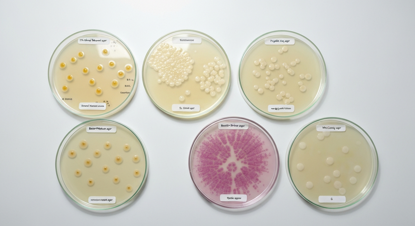
Staphylococcus aureus grows on a wide range of agars, but the ones you will encounter most often in a classroom or clinical context are blood agar, tryptic soy agar (TSA), mannitol salt agar (MSA), and Baird-Parker agar. Blood agar and TSA are general-purpose media that support robust growth with no special tricks. MSA and Baird-Parker are selective and differential, meaning they are designed to favor S. aureus over other bacteria and give you visual clues about its identity at the same time. If a medium contains bile salts or dyes that block Gram-positive bacteria, such as MacConkey or EMB agar, S. aureus will typically not grow at all.
What agar does Staphylococcus aureus grow on: key media

Marcus Holloway
25 Jul 2026
The biology behind S. aureus and why it matters for media selection
Before running through every agar on the list, it helps to understand what makes S. aureus tick, because the biology explains the media choices far better than memorization ever will. S. aureus is a Gram-positive coccus, which means it has a thick peptidoglycan cell wall on the outside of its cell membrane rather than a thin wall sandwiched between two membranes (the Gram-negative arrangement). That thick outer wall is chemically resistant to many dyes and bile salts that microbiologists deliberately use to block Gram-negative bacteria from growing. So when you see a medium advertised as 'selective for Gram-positives,' S. aureus generally sails right through that selection barrier.
S. aureus is also a facultative anaerobe, meaning it generates energy through aerobic respiration when oxygen is available but switches to fermentation when it is not. This metabolic flexibility means it is not picky about oxygen levels, and it will grow whether you incubate a plate in normal air or in a sealed container. On mannitol salt agar, this fermentation ability becomes visible as a color change in the medium, which we will get to shortly.
Perhaps the most practically important trait is halotolerance, the ability to survive and grow in high-salt environments. S. aureus grows comfortably at sodium chloride concentrations around 7.5% (the salt level used in MSA) and some strains can persist at concentrations even higher than that. Mechanistically, the bacterium copes with osmotic stress by pumping in compatible solutes, small molecules like glycine betaine and proline, that counterbalance the salt outside the cell without disrupting internal chemistry. This is why S. aureus is a recognized hazard in salty preserved foods, and it is exactly why MSA works as a selective medium: most other bacteria find that salt concentration lethal.
In terms of temperature and pH, S. aureus prefers conditions close to the human body. Its optimal growth temperature is roughly 35-37°C, though it can grow across a much wider range of about 7-48°C. Its optimal pH sits near neutral, around 6-7.5, though growth is possible across a surprisingly broad range from about pH 4 to pH 10 depending on other conditions. These tolerances explain why S. aureus can colonize the human skin and nasal passages so effectively, and why it shows up in improperly stored food that has been left at room temperature.
What agar actually does, and what makes media selective or differential
Agar itself is simply a structural scaffold. It is a polysaccharide extracted from red algae that dissolves in boiling water and sets into a firm gel when it cools. Bacteria cannot digest it (a key reason it replaced gelatin in the late 1800s), so it stays solid as colonies form on top. The nutrients, selective agents, and indicator dyes that determine what will grow, and what those colonies will look like, come from the other ingredients mixed in before the agar solidifies. There is a fuller discussion of this principle in the context of why bacteria grow on agar at all, which connects directly to foundational questions about what nutrients microorganisms actually need to reproduce.
Selective media use chemical weapons, things like antibiotics, bile salts, or high salt concentrations, to kill or inhibit organisms you do not want while allowing your target organism to grow. Differential media use indicator systems, usually pH-sensitive dyes or substrates that change color when metabolized, to distinguish one organism from another based on its biochemical behavior. Many of the most useful media for S. aureus are both selective and differential at the same time, giving you a two-step shortcut: first the organism survives the selective pressure, then its colony tells you something about its identity. Media that target Gram-positive organisms more broadly (selective Gram-positive agars like CNA/PEA) belong to this same family of thinking, and understanding how they work helps explain exactly where S. aureus fits on the spectrum of what grows and what does not.
General-purpose media: blood agar and TSA
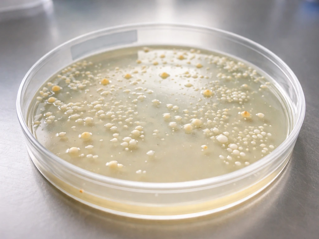
Blood agar (typically 5% sheep blood in a tryptic soy or Columbia agar base) is the workhorse of clinical microbiology. See our related guide on what bacteria grow on blood agar for a broader list of organisms and interpretations. It is non-selective, meaning most clinically relevant bacteria will grow on it, but it is rich enough to support fastidious organisms that need extra nutrients. S. aureus grows vigorously on blood agar, producing round, convex colonies that are typically golden-yellow or creamy white, ranging from about 1-3 mm in diameter after 18-24 hours at 35-37°C. On blood agar S. aureus colonies are often golden/yellow or white to cream and frequently show β‑hemolysis; small‑colony variants (SCVs) and injured/atypical strains may be non‑pigmented, reduced in size, and show weak or absent hemolysis, complicating visual identification Microbial Atlas / clinical culture images: Staphylococcus aureus on blood agar (descriptions of pigment/hemolysis and atypical forms). The golden pigment, produced by carotenoid compounds, gave the species its name: aureus is Latin for 'golden.' It is worth noting, though, that not every strain produces visible pigment, and small-colony variants (SCVs) or stressed cells can appear pale and surprisingly small, which is a common source of confusion in diagnostic labs.
Tryptic soy agar (TSA, sometimes called trypticase soy agar) and nutrient agar are similarly non-selective, nutrient-rich media that support excellent S. aureus growth. TSA is frequently used for subculturing, counting colonies, and preparing cultures for antibiotic susceptibility testing. Incubation at 35-37°C for 18-24 hours gives reliable, robust growth. These are the go-to media when you simply want S. aureus to grow without caring much about selective pressure or visual differentiation.
Selective and differential media for identifying S. aureus
Mannitol Salt Agar (MSA)
MSA is probably the first selective-differential medium most students encounter for S. aureus, and it is a nice demonstration of how salt tolerance and fermentation ability combine into a single diagnostic tool. The medium contains approximately 7.5% sodium chloride, which is toxic to most non-halotolerant bacteria, plus mannitol (a sugar alcohol) as a carbon source, and phenol red as a pH indicator. The logic is elegant: only salt-tolerant organisms survive to form colonies, and among those, organisms that ferment mannitol produce acid, which turns phenol red from its natural salmon-red color to yellow. S. aureus typically ferments mannitol, so you see yellow colonies surrounded by a yellow halo in the medium after 18-24 hours at 35-37°C.
One misconception worth addressing: the yellow color is a property of the medium reacting to acid, not necessarily the color of the colony itself. Colonies may look pale or cream-colored while the surrounding agar zone turns bright yellow. Both the colony color and the medium color change matter when you are reading a plate. Also important is that MSA is not perfectly specific for S. aureus. Some coagulase-negative staphylococci, such as S. haemolyticus and S. xylosus, also ferment mannitol and can produce yellow zones, leading to false positives. This is why MSA is a presumptive, not a definitive, identification tool. Confirmatory tests like the coagulase test, DNase test, or molecular methods are always recommended before calling an isolate S. aureus.
Baird-Parker Agar
Baird-Parker agar is the internationally recognized gold-standard medium for isolating and enumerating coagulase-positive staphylococci from food samples, and it is used in clinical settings as well. It contains potassium tellurite and lithium chloride as selective agents, plus egg-yolk emulsion as a differential substrate. S. aureus reduces potassium tellurite to elemental tellurium inside the cell, which produces the characteristic grey-black to jet-black colonies. The egg-yolk lecithinase reaction adds another layer: S. aureus produces lecithinase (and sometimes a lipase), causing a clear or opaque zone (halo) surrounding the black colony as the lecithin in the egg yolk is broken down. A classic S. aureus colony on Baird-Parker looks like a shiny black disc surrounded by a clear inner zone and sometimes an outer opaque ring. This combination of black coloration and the egg-yolk halo together form the presumptive identification signal, though confirmatory coagulase testing is still standard practice.
Chromogenic agars (CHROMagar Staph, SaSelect, and similar media)
Chromogenic agars represent a more modern approach. They incorporate colorless substrates that react with specific enzymes produced by the target organism, producing a visible pigment directly in or around the colony. CHROMagar Staph. aureus and similar products (SaSelect, Staph aureus ID) produce mauve, rose, or pink-mauve colonies for S. aureus based on enzyme activity specific to the species. These media can give a presumptive identification in as little as 18-24 hours and have reported sensitivities of 95% or higher in multiple independent evaluations. However, specificity can be more variable, particularly in specimens with diverse bacterial flora, where some studies report specificity in the 73-90% range depending on the formulation and the population tested. The practical takeaway is that chromogenic media are excellent screening tools, especially for rapid MRSA surveillance in clinical settings, but a positive result still warrants confirmation by phenotypic or molecular methods.
Selective Gram-positive agars: CNA and PEA
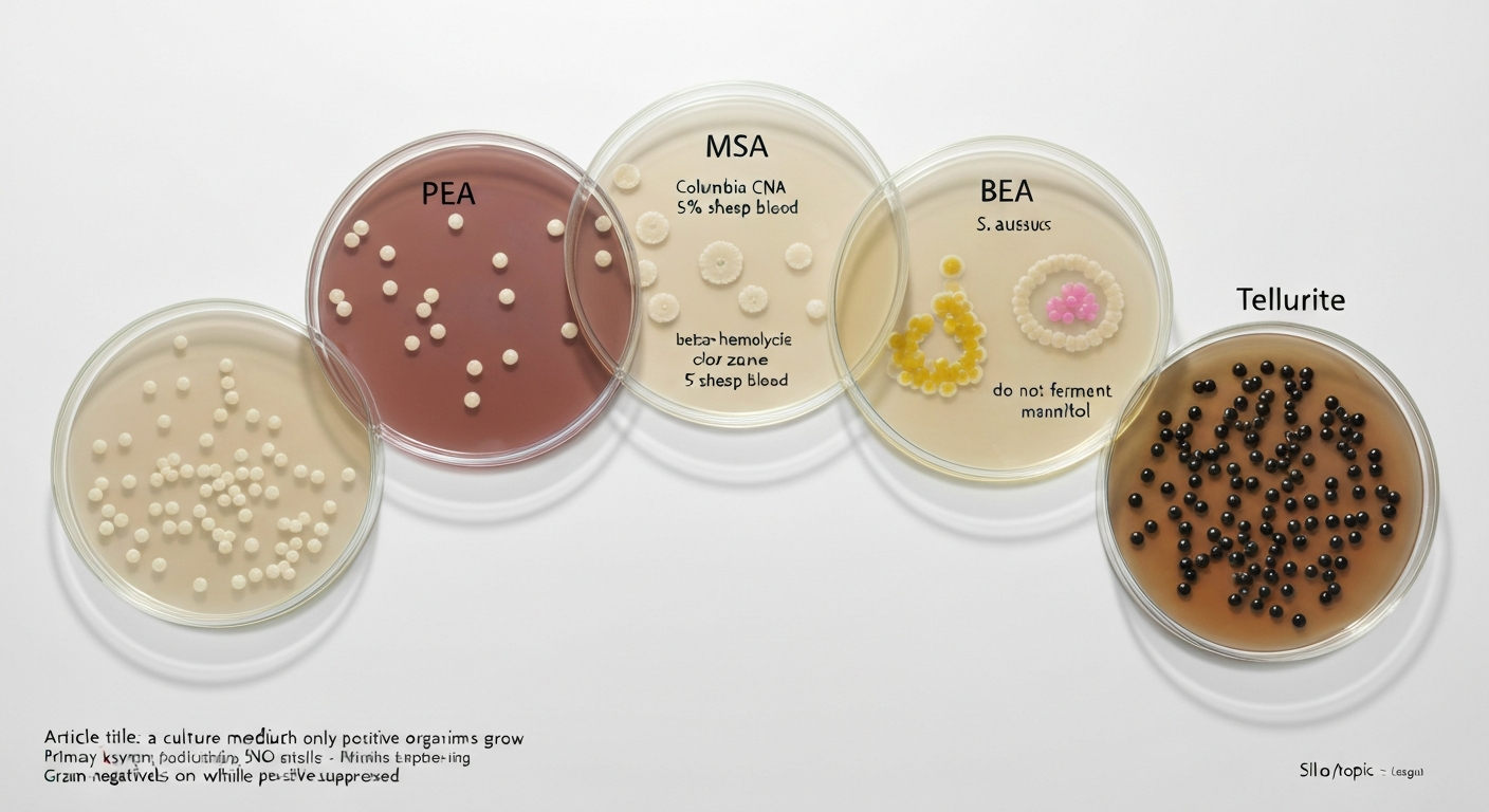
Columbia CNA agar (colistin-nalidixic acid blood agar) and phenylethyl alcohol agar (PEA) are designed to recover Gram-positive organisms from specimens likely to contain a mix of Gram-positive and Gram-negative bacteria. CNA contains colistin, which disrupts Gram-negative bacterial membranes, and nalidixic acid, a quinolone antibiotic that inhibits Gram-negative DNA replication. Together they block most Gram-negative bacilli while Gram-positive cocci like S. aureus grow freely. The blood component still allows hemolysis to be observed. PEA works by a different mechanism: phenylethyl alcohol disrupts the membranes of Gram-negative facultative anaerobes more than Gram-positives, achieving a similar selective effect. Both media are useful when you suspect S. For example, Columbia CNA agar and PEA are types of a culture medium on which only gram positive organisms grow. aureus in a sample that could be overgrown by Gram-negative organisms, such as certain wound or environmental samples.
Reading the plate: colony appearances and key reactions
Knowing which medium to use is only half the job. Reading and interpreting what you see is equally important, especially because a single organism can look quite different on different media. Here is a practical summary of the key visual cues.
| Medium | S. aureus colony appearance | Key differential reaction | Presumptive indicator of S. aureus? |
|---|---|---|---|
| Blood agar (5% sheep blood) | Golden-yellow or cream, 1-3 mm, round, convex | Beta-hemolysis: clear zone of complete red blood cell lysis around colony | Presumptive (confirm with coagulase) |
| Tryptic soy agar (TSA) / Nutrient agar | Golden-yellow or cream, similar morphology | None (non-differential) | No differential information |
| Mannitol Salt Agar (MSA) | Yellow or cream colonies; surrounding agar turns yellow | Mannitol fermentation: phenol red indicator turns yellow (acidification) | Presumptive (some CoNS also ferment mannitol) |
| Baird-Parker agar | Grey-black to jet-black, shiny, 1.5-2.5 mm | Clear/opaque egg-yolk lecithinase halo surrounding black colony | Presumptive (confirm with coagulase) |
| CHROMagar Staph / SaSelect | Mauve, rose, or pink-mauve colonies | Chromogenic enzyme reaction specific to S. aureus | Presumptive (high sensitivity; confirm positives) |
| Columbia CNA agar | As on blood agar; beta-hemolysis visible | Gram-negatives inhibited; Gram-positives grow normally | Growth context only (not differential for S. aureus specifically) |
Beta-hemolysis on blood agar is one of the most recognized S. aureus signatures, but it is important not to overinterpret it. Beta-hemolysis means the organism produces hemolysins (toxins) that completely lyse red blood cells, leaving a transparent zone around the colony. Not every S. aureus strain produces strong beta-hemolysis, and some strains show weak, incomplete (alpha) hemolysis or no visible hemolysis at all, particularly small-colony variants or strains from certain clinical contexts. Conversely, other species, including some streptococci, also produce beta-hemolysis. So hemolysis on blood agar is a useful clue, not a conclusion, and the coagulase test (tube or latex agglutination) remains the definitive phenotypic confirmation step for S. aureus.
Media that inhibit S. aureus and why
MacConkey agar and eosin methylene blue (EMB) agar are two of the most common media that will inhibit S. aureus growth, and understanding why helps reinforce what makes Gram-positive bacteria fundamentally different from Gram-negative ones. MacConkey agar contains bile salts and crystal violet dye, both of which disrupt the cell membrane of Gram-positive bacteria. The Gram-positive cell wall, despite being thicker in terms of peptidoglycan, does not have the outer membrane that shields Gram-negative bacteria from these detergent-like molecules. EMB agar uses eosin and methylene blue dyes that are similarly inhibitory to Gram-positive organisms. The result is that S. aureus either fails to grow at all on these media or grows very poorly, making them completely inappropriate for primary S. aureus isolation.
More generally, any medium that relies on antibiotics selectively toxic to Gram-positive bacteria (such as vancomycin, which targets peptidoglycan synthesis, at high concentrations used for Gram-negative enrichment) or that uses highly alkaline or acidic conditions outside S. aureus tolerance ranges will suppress its growth. This is why understanding the fundamental biology of the organism always comes first when choosing media: the chemistry of the medium has to match, or deliberately mismatch, the biology of what you are trying to grow.
S. aureus compared to streptococci and Gram-negative organisms
Students often ask how S. aureus differs from streptococci on culture media, since both are Gram-positive cocci that show up in similar clinical specimens. There are meaningful differences that are worth knowing. Both grow on blood agar, but streptococci generally require richer, more specific nutritional supplementation in some cases and will not grow on MSA because they lack the halotolerance that S. aureus and other staphylococci possess. On blood agar, the hemolysis patterns differ: S. pyogenes (Group A Streptococcus) also produces beta-hemolysis, which can create confusion, but the colony morphology is different (streptococcal colonies tend to be smaller and more translucent), and streptococci are catalase-negative while S. aureus is catalase-positive. That single catalase test, dropping hydrogen peroxide on a colony and watching for bubbles, is a quick way to separate the two genera. Questions about which specific media Streptococcus will and will not grow on connect directly to how media selectivity differs between these two important genera of Gram-positive cocci.
Gram-negative organisms like Escherichia coli or Pseudomonas aeruginosa present an entirely different growth profile. They thrive on MacConkey and EMB, media that kill or inhibit S. aureus. On blood agar, both Gram-positive and Gram-negative organisms can grow, which is why blood agar is described as nonselective. For a concise guide to which media support both groups, see most gram positive and gram negative organisms will grow on. This is the exact contrast captured in the broader discussion of which media support both Gram-positive and Gram-negative organisms versus those designed to select one group over the other. S. aureus will not grow on media designed exclusively for Gram-negative isolation, and the key structural reason always comes back to that Gram-positive cell wall chemistry.
| Feature | S. aureus | Streptococcus spp. | Gram-negative rods (e.g., E. coli) |
|---|---|---|---|
| Gram stain | Gram-positive cocci in clusters | Gram-positive cocci in chains or pairs | Gram-negative rods |
| Catalase test | Positive (bubbles with H2O2) | Negative (no bubbles) | Positive (most) |
| Blood agar growth | Yes, robust; often beta-hemolysis | Yes; alpha, beta, or gamma hemolysis by species | Yes (non-selective) |
| Mannitol Salt Agar (7.5% NaCl) | Yes (mannitol fermentation, yellow zone) | No (salt-sensitive) | No (salt-sensitive + bile salt inhibition) |
| MacConkey / EMB agar | No (inhibited by bile salts/dyes) | No (inhibited) | Yes (designed for Gram-negatives) |
| Baird-Parker agar | Yes; black colonies with egg-yolk halo | Inhibited or very poor growth | Inhibited |
| CNA / PEA agar | Yes (Gram-positive selective) | Yes (Gram-positive selective) | Inhibited |
A note on biofilms and growth beyond the plate
Most of this article focuses on planktonic growth, where individual bacteria multiply as free-floating or surface-sitting cells on an agar plate. It is worth knowing that S. aureus is also a capable biofilm former, meaning it can attach to surfaces and encase itself in a protective matrix of polysaccharides and proteins. Biofilm growth does not change which media S. aureus will grow on in a standard culture sense, but it is relevant to food safety, medical device contamination, and antibiotic resistance, since bacteria within biofilms can be dramatically harder to kill than their planktonic counterparts. Understanding the distinction between colony growth on agar and biofilm formation in real environments is part of a complete picture of how S. aureus behaves in the world.
Safety and context for working with S. aureus
S. aureus is classified as a Biosafety Level 2 (BSL-2) organism, meaning work with it requires standard microbiological practices, appropriate personal protective equipment, and access to a properly equipped laboratory. It is not a microorganism for uncontrolled experimentation at home. This article is written to help students and educators understand the principles behind media selection and bacterial growth, not to provide a step-by-step culturing protocol. If you are conducting laboratory exercises, always follow your institution's safety guidelines and your instructor's directions, and never attempt to culture clinical or environmental isolates outside of a properly supervised setting.
Quick reference: which agars S. aureus grows on
To bring everything together in one place, here is a concise overview of how S. aureus behaves across the most common microbiological media you are likely to encounter in a classroom or clinical lab setting.
- Blood agar (5% sheep blood): grows well; golden-yellow or cream colonies; often beta-hemolysis (clear zone around colony)
- Tryptic soy agar (TSA) / Nutrient agar: grows robustly; used for subculture and susceptibility testing; no differential reaction
- Mannitol Salt Agar (MSA): grows well due to halotolerance; ferments mannitol to produce acid, turning phenol-red medium yellow; presumptive for S. aureus
- Baird-Parker agar: grows well; grey-black to black colonies due to tellurite reduction; egg-yolk lecithinase halo; internationally used for food safety testing
- CHROMagar Staph / SaSelect and similar chromogenic media: grows well; produces mauve/rose/pink colonies via specific enzyme reactions; high sensitivity, variable specificity
- Columbia CNA agar / PEA agar: grows well; Gram-negative organisms inhibited; blood component allows hemolysis observation
- MacConkey agar: does not grow (inhibited by bile salts and crystal violet)
- EMB agar: does not grow (inhibited by eosin and methylene blue dyes)
FAQ
Quick answer: What agar does Staphylococcus aureus grow on?
S. aureus grows well on general-purpose agars (blood agar, tryptic soy agar/nutrient agar), halotolerant selective media (Mannitol Salt Agar), selective/differential plates for staphylococci (Baird‑Parker agar), and chromogenic S. aureus or MRSA media (CHROMagar Staph. aureus, SaSelect). It also grows on Gram‑positive selective plates (e.g., Columbia CNA with 5% sheep blood). Media formulated to inhibit Gram‑positives (MacConkey, EMB, and other bile‑salt/dye containing media) typically prevent its growth.
Which general-purpose media support S. aureus growth and what do colonies look like?
Blood agar (5% sheep blood) and tryptic soy agar (TSA)/nutrient agar support robust growth. Typical colonies are round, smooth, often golden‑yellow to cream (staphyloxanthin pigment) and commonly show β‑hemolysis (clear zones) on blood agar after 18–24 h at 35–37°C. Atypical or small‑colony variants may be nonpigmented, smaller, and show weak or absent hemolysis.
What does Mannitol Salt Agar (MSA) do and how does S. aureus appear on it?
MSA is selective (high NaCl ≈7.5% selects for staphylococci) and differential (phenol red indicator detects mannitol fermentation). S. aureus typically ferments mannitol, acidifying the medium and turning the agar yellow; colonies may appear yellow or pale with a surrounding yellow zone. Note: some coagulase‑negative staphylococci can also ferment mannitol, so MSA is presumptive, not definitive.
What is Baird‑Parker agar and how does S. aureus behave on it?
Baird‑Parker agar contains tellurite and egg‑yolk; tellurite is reduced by staphylococci to elemental tellurium producing grey‑black to black colonies, and egg‑yolk lecithinase activity often yields a clear or opaque halo. It is widely used as a selective/differential medium for coagulase‑positive staphylococci (including S. aureus) especially in food testing.
What are chromogenic S. aureus plates and how reliable are they?
Chromogenic media contain substrates and dyes that produce species‑specific colony colors (e.g., mauve/rose for S. aureus). They speed presumptive identification and often have high sensitivity (commonly ≥95%) but specificity varies with formulation and local flora; positive results should be confirmed by phenotypic or molecular tests when clinical or regulatory decisions depend on accuracy.
Which agars typically inhibit or prevent growth of S. aureus and why?
MacConkey and EMB agars (and other bile salt/dye‑containing media) are formulated to inhibit Gram‑positive organisms using bile salts, crystal violet, eosin, or methylene blue; these components disrupt Gram‑positive cell walls or selective physiology, so S. aureus generally does not grow or grows poorly on them. Media containing antibiotics that target Gram‑positives will also inhibit S. aureus.