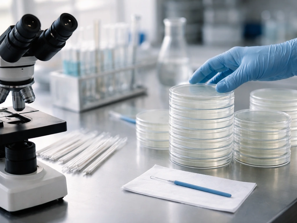
Microbiologists grow bacteria because you cannot study what you cannot see, and you cannot see much from a handful of invisible cells. Culturing bacteria, which means giving them the right conditions to multiply into visible colonies, is how researchers learn what those organisms need to survive, how to identify them, how to kill them, and how they behave when conditions shift. It is the foundational move that makes almost every downstream experiment possible, from diagnosing an infection to testing whether an antibiotic will work. CDC biosafety training notes that BSL-3 builds on BSL-2 containment and gives Mycobacterium tuberculosis as a typical example of a microbe worked with under BSL-3 blank" rel="noopener noreferrer">CDC LC Quick Learn.
Why Would Microbiologists Want to Grow Bacteria? Uses Explained

The core reasons microbiologists culture bacteria
There is no single reason microbiologists grow bacteria. There are at least four, and they are tightly linked. First, culturing is how you figure out what conditions a bacterium needs to flourish, because growth (or the lack of it) is the feedback. Second, it is the starting point for identification, since you need isolated colonies before you can run biochemical tests, microscopy, or molecular characterization. Third, controlled growth lets researchers probe physiology and genetics, watching how gene expression and metabolism shift when you change temperature, pH, oxygen, or nutrients. Fourth, growing bacteria under defined conditions is essential for applied goals like testing antibiotic susceptibility, monitoring contamination in hospitals or water systems, and supporting food safety investigations.
What ties all four together is the underlying biology. Bacteria do not grow the same way in every environment. They flourish or stay dormant depending on a specific combination of conditions, and the only way to map that combination is to deliberately vary it and watch what happens. That is what culturing is really for.
How growing bacteria reveals what they actually need
One of the most direct uses of bacterial culture is learning the growth requirements of a given species. You do this by changing one condition at a time and measuring whether colonies form, how fast, and how robustly. The major variables are temperature, pH, oxygen availability, water activity, and nutrient sources, and they are interconnected rather than independent levers.
Temperature, pH, and oxygen
Temperature affects how quickly bacterial enzymes work and whether proteins stay folded. Most human pathogens are mesophiles, meaning they grow best around 37°C, which is human body temperature. That is not a coincidence. pH matters because bacteria must maintain stable internal acidity even when the outside environment swings acidic or alkaline.
When external pH drops, bacteria activate proton-pumping pathways to prevent their cytoplasm from acidifying, and if the stress is too great, growth stops. Oxygen is equally defining. Aerobic bacteria use oxygen as the final electron acceptor in respiration. Obligate anaerobes cannot tolerate it at all.
Many clinically important bacteria are facultative anaerobes, meaning they can switch strategies depending on what is available. Laboratories replicate this by incubating cultures under aerobic, microaerophilic, or fully anaerobic atmospheres, and a mismatch between the organism's oxygen strategy and the incubation conditions produces a false negative, meaning no growth even though viable cells are present.
Moisture and nutrients
Water activity (written as aw) measures how much free water is available for biological use. Most bacteria thrive at aw near 0. 99, which is close to pure water, and growth shuts down at sufficiently low a_w values, around 0. 6 for many species, because the osmotic stress becomes too great.
This is exactly why drying and salting are ancient food preservation techniques. Nutrients round out the picture. Bacteria need carbon and nitrogen sources to generate the reducing equivalents that power metabolism, whether through aerobic respiration, anaerobic respiration, or fermentation. Different species have very different nutritional requirements, and the choice of growth medium in the lab directly reflects this.
Understanding these needs through culture is the same foundational question as asking why germs grow where they do in the environment.
What growth phases tell you
When bacteria are transferred into fresh medium, they go through predictable phases: a lag phase where they ramp up metabolic machinery, an exponential (log) phase where cells divide at their maximum rate, a stationary phase when nutrients run low and waste builds up, and a death phase. Watching how quickly a culture moves through these stages under different conditions tells you a great deal about what that organism finds stressful.
Watching how quickly a culture moves through these stages under different conditions tells you a great deal about what that organism finds stressful the stage in which microorganisms grow and reproduce. A prolonged lag phase often means the conditions are suboptimal. A slow exponential rate can point to a missing nutrient or a pH that is slightly off.
And some bacteria, under severe stress like starvation or sharp pH swings, enter a viable but nonculturable (VBNC) state, where they are metabolically active but stop forming colonies on standard media. VBNC is a real complication for interpretation because no colonies does not always mean no bacteria.
Using cultures to identify and characterize bacteria
Before you can identify a bacterium, you need a pure culture, which means isolated colonies of a single organism. Clinical labs get there by inoculating patient specimens onto several types of media at once and incubating under different atmospheric conditions to capture whatever pathogens might be present. The three major media categories are selective (which suppress unwanted organisms while allowing the target to grow), differential (which make different organisms look visually distinct based on a biochemical reaction), and enrichment (which boost the growth of organisms present in low numbers without necessarily suppressing others).
A classic example is MacConkey agar. It is both selective, inhibiting Gram-positive bacteria, and differential, turning lactose-fermenting colonies pink while non-fermenters stay colorless. That color difference is not cosmetic. It reflects underlying metabolic chemistry and immediately narrows down what you are looking at. Chromogenic media take this further by linking enzyme activity to colorimetric reactions, allowing presumptive identification from colony color alone, though these have specificity limitations depending on the organism and specimen complexity.
Once isolated, colonies are characterized by their morphology (size, shape, color, texture, surface appearance), microscopic appearance after staining, the atmosphere and time required for growth, and biochemical or physiological traits. In modern labs, antigenic testing (serotyping) and nucleotide sequence analysis are added on top. But all of that downstream work depends on having a pure, well-grown colony to start from. Culture is the amplification step that makes a microscopic population large enough to work with.
Studying physiology and genetics under controlled conditions
Controlled culture conditions are essential for studying how bacteria work at the cellular and molecular level. When you grow the same strain under two different conditions and compare the results, you are essentially running an experiment on what the organism needs and how it adapts. This is where growth biology connects directly to genetics.
Bacteria regulate gene expression in response to environmental signals through systems like sigma factors and two-component signaling pathways. Sigma factors are proteins that direct RNA polymerase to specific genes. The housekeeping sigma factor drives bulk transcription during normal growth, while alternative sigma factors kick in under stress, shifting the cell's priorities. Two-component systems work by sensing an environmental signal outside the cell, then relaying that signal inward to change which genes get turned on.
These are not abstract mechanisms. They are the reason a bacterium behaves differently at pH 5 than at pH 7, or at 25°C versus 37°C. Culturing under deliberately varied conditions, and then comparing gene expression, enzyme activity, or metabolic output, is how researchers map these regulatory circuits.
Understanding microbial growth phases is also central here. Researchers studying stress responses often need bacteria in a specific phase, because a cell in log phase expresses very different genes than one in stationary phase. Controlling the culture means controlling which biological state you are actually observing.
Applied benefits: treatments, contamination, and food and water safety
Growing bacteria is not only about basic science. It has direct practical consequences in medicine, public health, and food safety.
Antimicrobial susceptibility testing
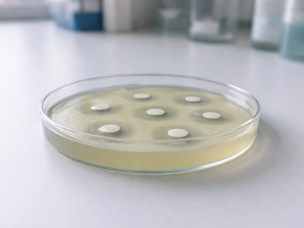
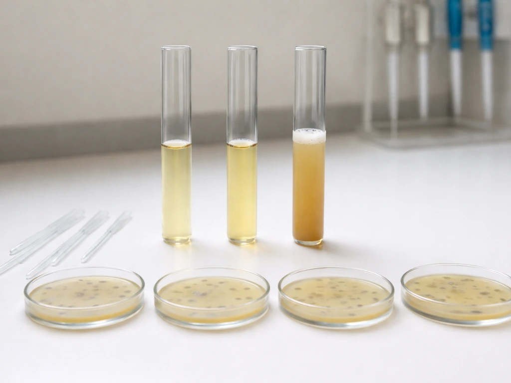
To find out whether an antibiotic will work against a particular bacterial isolate, you need to grow that isolate under controlled conditions and expose it to the drug. Disk diffusion testing places antibiotic-impregnated disks on a plate inoculated with bacteria. A clear zone (called a zone of inhibition) around the disk means the antibiotic is suppressing growth at that concentration. Broth microdilution testing, standardized under protocols like CLSI M07, measures the minimum concentration of antibiotic needed to prevent visible growth in liquid medium. Without culture, you cannot do either test. Resistance surveillance programs like the CDC's NARMS (National Antimicrobial Resistance Monitoring System) depend on culturing isolates and running susceptibility panels to track which antibiotics are losing effectiveness.
Contamination monitoring
In healthcare settings, environmental culturing involves swabbing surfaces or collecting water samples and growing whatever microorganisms are present on standard recovery media like tryptic soy agar (TSA). In microbiology, the site where pathogens grow is called the incubation environment. This tells infection control teams whether cleaning protocols are working. Water sampling has a practical wrinkle worth knowing: residual disinfectants like chlorine can suppress growth in the sample even after collection, so water samples are often collected into sodium thiosulfate to neutralize the chlorine before culturing. Missing that step produces false negatives, which is exactly the kind of result-interpretation pitfall that matters when you are trying to declare a water system safe.
Food and water safety investigations
Food safety microbiology relies heavily on selective and enrichment media to detect specific pathogens at low concentrations in complex matrices. The same growth biology principles apply here. A food with low water activity inhibits bacterial growth, which is why dried goods store longer. A food at neutral pH in warm, moist conditions is a much higher-risk environment. Culture-based methods let investigators confirm whether a food sample actually contains viable, colony-forming pathogens, which is relevant both for outbreak investigations and for routine monitoring programs.
What to observe and how to interpret growth results safely
If you are learning to interpret bacterial growth experiments, whether in a classroom or a basic lab setting, there are a few principles worth keeping in mind.
- Match your incubation conditions to the organism's known requirements. If you are working with a facultative anaerobe but incubating in full aerobic conditions, you may still get growth, but you may miss something important about its behavior under oxygen-limited conditions. Oxygen mismatches are one of the most common sources of false negatives.
- Know what your medium is designed to do. A selective medium that suppresses Gram-positives will not tell you whether Gram-positives are absent from your sample. It just means you chose to ignore them. Always think about what your medium allows versus what it suppresses.
- Interpret no growth carefully. No visible colonies does not automatically mean no bacteria. VBNC cells remain metabolically active but stop forming visible colonies when they are stressed by starvation, extreme temperature, pH swings, osmotic stress, or disinfectants. If your sample came from a harsh environment, a negative plate result deserves skepticism.
- Watch colony morphology closely. Colony size, color, texture, surface appearance, and the presence of multiple distinct colony types are all informative. Multiple morphologies on a plate suggest a mixed culture, which means you need to subculture onto fresh plates to isolate individual strains before drawing any identification conclusions.
- Track growth phases over time. When you are trying to understand growth conditions, a single time point is rarely enough. Photographing or measuring at multiple intervals shows you whether you are seeing a genuine lag, a slow exponential, or early stationary phase, which tells you a lot about whether the conditions are optimal.
- Follow appropriate biosafety practices. In any lab context, unknown bacteria are handled following BSL-2 standard precautions as a baseline, treating all biological specimens as potentially hazardous. If working with known or suspected high-risk pathogens, BSL-3 protocols with additional containment apply. When in doubt, follow your institution's biosafety guidelines rather than improvising.
The deeper takeaway is that growing bacteria is an act of asking questions. Every condition you set, every medium you choose, every time point you observe, is a question about what the organism needs and how it responds. That is why understanding the biology of microbial growth, the roles of temperature, pH, oxygen, moisture, and nutrients, is not just background knowledge. It is the framework that makes all the results interpretable. The conditions where microorganisms grow best are the same conditions that culture experiments are designed to explore, and tracing that logic through to applied outcomes like antibiotic testing or contamination monitoring is exactly what makes microbiology so practically powerful.
FAQ
What is the difference between “growing bacteria” and simply detecting their DNA or RNA?
Culture shows whether cells can form colonies under specific conditions, which is closer to viability and growth potential. Molecular detection can find genetic material even when cells are dead or in a nonculturable state, so it does not always answer whether the organism would actually multiply in your environment.
Why do some cultures stay negative even when a test is otherwise suspected to be positive?
A common cause is mismatch between incubation conditions and the bacterium’s strategy (for example, oxygen level or temperature). Another cause is viable but nonculturable (VBNC) cells, where metabolism continues but standard media and times do not produce colonies.
How do microbiologists decide which culture medium is the “right” one?
They usually start by narrowing the target group, then choose medium type based on that goal. Selective media suppress likely competitors, differential media help sort phenotypes by a biochemical signal, and enrichment media increase the chance of recovering low-abundance organisms from complex samples.
How does the choice of incubation time affect conclusions about growth?
Many bacteria grow at different speeds, so too-short incubation can create false negatives, and too-long incubation can overgrow slower organisms or let stressed cells recover. Interpreting results usually requires using organism- and test-specific incubation windows.
Why is “pure culture” emphasized before identification work?
Mixed colonies can produce misleading colony morphology and conflicting biochemical signals, especially when multiple bacteria grow with similar appearance. Isolation into a single-colony origin ensures downstream tests reflect one organism’s traits rather than a blend.
What are the practical limits of using colony counts to estimate how many bacteria are in a sample?
Colony forming units depend on whether cells are able to separate and grow as individual colonies. Clumping, stress, or VBNC behavior means the CFU count can underestimate total bacterial cells present in the original sample.
Why would a water or environmental sample need special collection handling before culturing?
Residual disinfectants can keep killing cells after sampling, reducing recovery and producing false negatives. Neutralizing agents like sodium thiosulfate are used to stop disinfectant activity so the culture reflects what was in the water at collection time.
How do microbiologists avoid false susceptibility results when testing antibiotics?
They must use standardized growth conditions and inoculum preparation, since growth state strongly affects antibiotic effect. Testing an isolate from the wrong growth phase, or using nonstandard incubation, can shift the apparent zone size or minimum inhibitory concentration.
What’s the safest way to think about growth phases when interpreting experiments?
Culture phase matters because gene expression and stress responses change across lag, log, stationary, and death phases. If your goal is to study regulation tied to active growth, comparing log-phase cells is often more informative than comparing stationary-phase or mixed-phase cultures.
In classrooms or introductory labs, why might the incubation atmosphere be the key variable?
Because oxygen strategy (aerobic, facultative, or obligate anaerobic) determines whether a bacterium can respire and grow. Incubating with the wrong atmosphere can make viable cells fail to form colonies, leading to confusion about media effectiveness or organism presence.