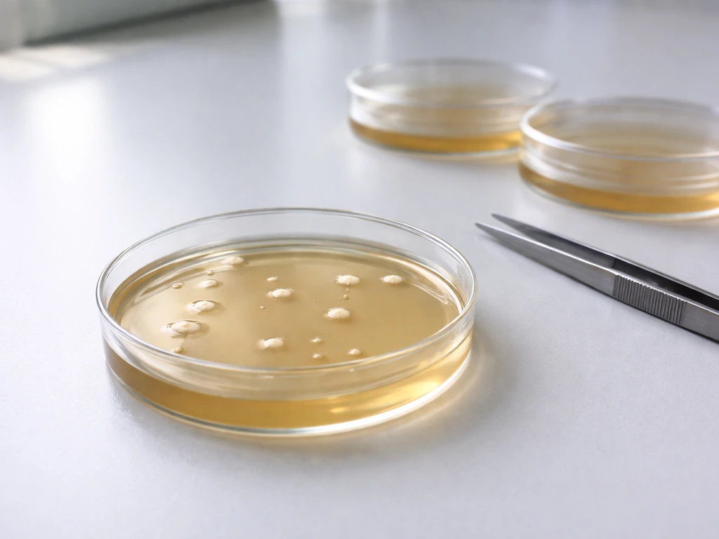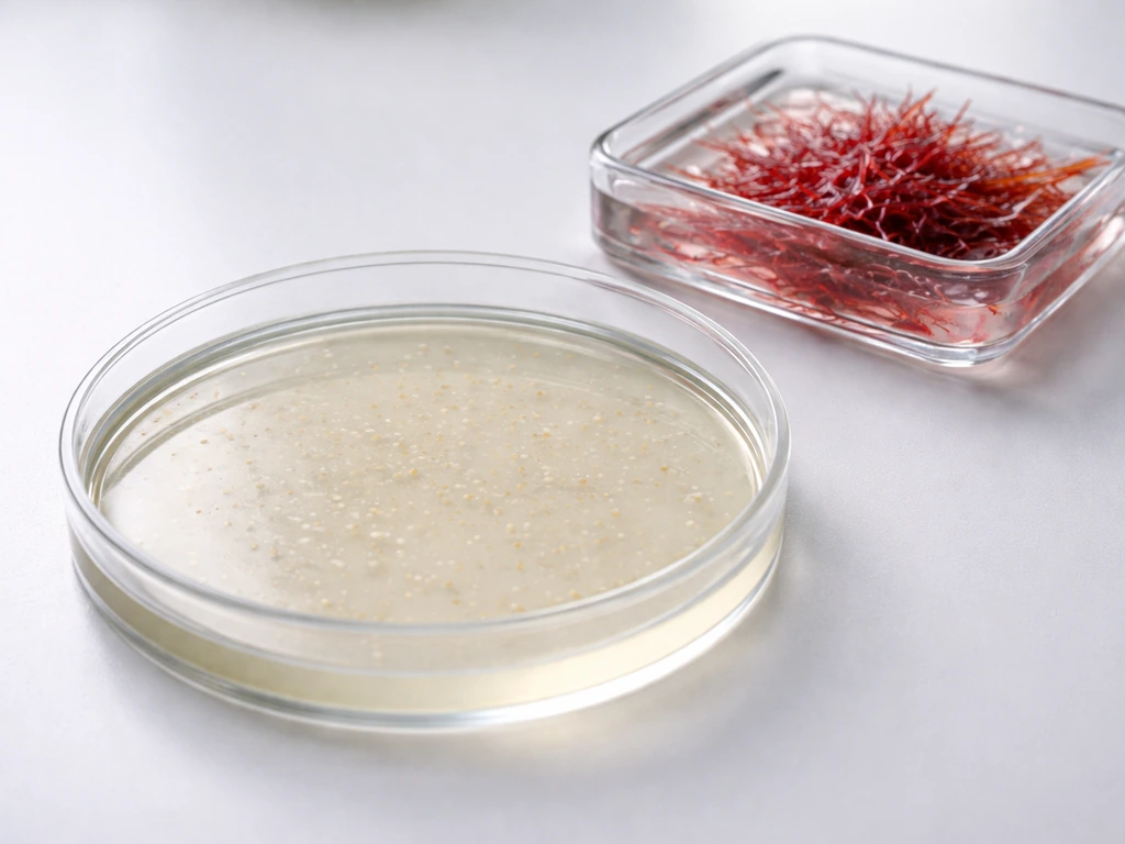
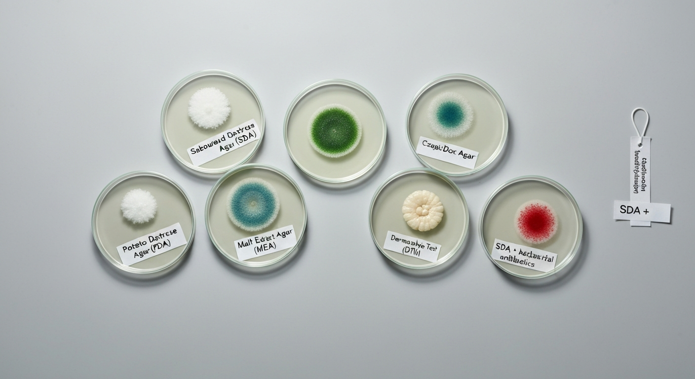
The most widely used agar for growing fungi is Sabouraud Dextrose Agar (SDA). Its acidic pH of around 5.6 and high glucose concentration (40 g/L) create conditions that favor fungi while discouraging most bacteria, making it the default starting point in mycology labs, classrooms, and diagnostic settings worldwide. Beyond SDA, Potato Dextrose Agar (PDA), Malt Extract Agar (MEA), and defined media like Czapek-Dox agar each serve specific purposes depending on the fungal group being studied, the question being asked, and whether selectivity or open-ended growth is the goal.
What Agar Is Used to Grow Fungi: Common Media & Uses

Marcus Holloway
12 Jul 2026
What agar actually is and why it works for fungi
Before getting into specific media, it helps to understand what agar is and why it became the standard solidifying agent in microbiology. Agar is a polysaccharide extracted from red seaweeds, primarily Gelidium and Gracilaria species. It is composed mainly of two fractions: agarose (the gelling component) and agaropectin (a sulfated, charged fraction). The reason agar became so useful is that it forms a thermo-reversible gel with a large and very deliberate gap between its melting and setting temperatures. Agar gels on cooling at roughly 32 to 45 °C and only melts again when heated to about 85 to 95 °C. That gap is critical. It means you can pour liquid agar into plates at around 45 to 50 °C without it solidifying too quickly, and once the plates are poured and set, they stay solid even inside a 30 °C incubator for weeks. For a concise comparison of how agar supports both fungal and bacterial growth, see why is agar used to grow bacteria.
Agar is also chemically inert in the sense that most fungi and bacteria cannot break it down easily. It forms a stable, porous gel at concentrations as low as 1.5% (w/v), which provides a physical surface for colonies to grow on without itself becoming a significant nutrient source. This inertness matters enormously: you can add whatever nutrients you want to agar-based media without worrying that the gelling agent will interfere. The agar is essentially just the scaffold. The nutrients, pH adjusters, selective inhibitors, and indicator dyes are where the real biological action happens.
Who uses different fungal media, and for what purpose
Not every situation calls for the same medium, and understanding who is doing the culturing and why shapes every media decision. Students working at the introductory level are typically using fungal media to observe colony morphology, study sporulation, or compare growth rates across organisms. For these purposes, PDA and MEA are friendly choices because they support vigorous, visually rich growth and are widely available in school lab supply catalogs.
Educators designing classroom investigations need media that are both effective and safe to handle. Non-selective media (without antibiotics or cycloheximide) reduce chemical handling concerns and are appropriate when work is done with known, low-risk fungal species under proper supervision. For anyone going further into diagnostic or clinical applications, such as a college-level microbiology course or a professional lab context, SDA with selective antibacterial supplements becomes the workhorse. Diagnostic lab technicians processing clinical specimens (skin scrapings, nail clippings, hair) typically inoculate both a selective and a non-selective medium in parallel to avoid missing organisms that selective inhibitors might suppress. For dermatophyte specimen processing, laboratories should inoculate both selective (e.g., Mycobiotic/DTM/Mycosel with cycloheximide+antibiotics) and nonselective media (e.g., SDA, PDA without cycloheximide) because cycloheximide can inhibit certain pathogens and because selective media can produce false positives on DTM (non‑dermatophytes may also change indicator color) Clinical guidelines recommend inoculating both selective (e.g., Mycobiotic/DTM/Mycosel with cycloheximide+antibiotics) and nonselective media (e.g., SDA, PDA without cycloheximide) because cycloheximide can inhibit certain pathogens and selective media can produce false positives on DTM..
A catalog of common fungal media and how to read a formulation
Every microbiological medium is essentially a recipe, and reading a formulation tells you immediately what biological question it was designed to answer. The key variables are: the carbon source (what the fungus burns for energy), the nitrogen source (what it uses to build proteins and nucleic acids), the pH, and any selective or differential additives. Most fungal media use glucose or sucrose as the primary carbon source, organic nitrogen from peptones or malt extract, and an acidic pH to favor fungi over bacteria. Selective media go further by adding antibacterial agents to kill contaminating bacteria, or antifungals like cycloheximide to suppress fast-growing mold contaminants. Here is a comparative overview of the major fungal media you are likely to encounter.
| Medium | Primary carbon source | Nitrogen source | Approximate pH | Key selective features | Main uses |
|---|---|---|---|---|---|
| Sabouraud Dextrose Agar (SDA) | Dextrose (glucose) 40 g/L | Peptones ~10 g/L | ~5.6 | Acidic pH, high glucose; optional antibacterial supplements | General fungal isolation, yeasts and molds, dermatophytes |
| Potato Dextrose Agar (PDA) | Glucose 20 g/L + potato extract 4 g/L | Potato extract (trace) | ~5.6 (can be acidified to 3.5) | Mild selectivity; acidic pH; can support bacteria | General fungal culture, sporulation studies, food/environmental mycology |
| Malt Extract Agar (MEA) | Malt extract ~20–30 g/L | Malt extract + optional peptone ~5 g/L | ~5.4–5.5 | Slightly acidic; rich in sugars and vitamins | Saprophytic fungi, yeasts, sporulation, food spoilage organisms |
| Czapek-Dox Agar | Sucrose 30 g/L | Sodium nitrate 3 g/L (inorganic) | ~7.2 | Defined medium; only inorganic nitrogen | Aspergillus, Penicillium taxonomy, physiological studies |
| Dermatophyte Test Medium (DTM) | Dextrose 10 g/L | Soy peptone 10 g/L | ~5.5 | Cycloheximide + chloramphenicol + gentamicin + phenol red indicator | Clinical dermatophyte detection, selective and differential |
Sabouraud Dextrose Agar: the standard reference point
SDA was developed by French dermatologist Raymond Sabouraud in the late 19th century specifically for cultivating dermatophytes, the fungi responsible for ringworm, athlete's foot, and nail infections. Its formulation has remained relatively stable ever since. A standard commercial preparation dissolves at 65 g/L dehydrated powder to give approximately 10 g/L peptone (often split between two peptone sources), 40 g/L dextrose (glucose), and 15 g/L agar, with a final pH of around 5.6.
The high glucose concentration serves two roles. First, glucose is the preferred carbon and energy source for most fungi, fueling rapid growth. Second, at 40 g/L, glucose slightly lowers the water activity of the medium, creating a mildly osmotic environment that fungi handle better than most bacteria. This is not a fully selective environment on its own, but combined with the acidic pH of 5.6, it meaningfully tilts the balance. Most common bacteria grow optimally at pH 6.5 to 7.5. At pH 5.6, their growth slows or stops altogether while fungi, which often prefer slightly acidic conditions, continue growing comfortably.
When labs need stronger selectivity, particularly when processing specimens from the skin or environment where bacterial contamination is expected, SDA is supplemented with filter-sterilized antibacterial agents added aseptically to cooled agar at about 45 to 50 °C. Chloramphenicol (a broad-spectrum bacteriostatic agent) and gentamicin (effective primarily against Gram-negative bacteria) are the most common choices. These are added after autoclaving, not before, because autoclaving would degrade the antibiotic activity.
SDA's main limitations are its lack of differential capability (it does not tell you what type of fungus is growing, only that something is growing) and the fact that at pH 5.6 without cycloheximide, fast-growing saprophytic molds can still overgrow slower pathogens. That is why specialized selective formulations like Mycosel or Mycobiotic agar add cycloheximide to SDA to suppress saprophytes. However, cycloheximide is a eukaryotic protein synthesis inhibitor that can also suppress some clinically important fungi, including certain Cryptococcus species and some dimorphic pathogens, so using it as the only medium is not recommended in clinical settings.
Potato Dextrose Agar: versatile and visually rich
PDA has a simpler and more natural origin story. The standard Oxoid/Thermo Fisher formulation calls for potato extract at 4 g/L (equivalent to infusing 200 g of fresh potato), glucose at 20 g/L, and agar at 15 g/L, with a native pH of about 5.6. The potato extract contributes trace nitrogen, vitamins, and a mix of organic compounds that fungi find very hospitable. The glucose concentration is lower than in SDA (20 g/L vs 40 g/L), but that actually suits many saprophytic fungi and plant pathogens that do not need the extreme osmotic challenge of SDA.
PDA is especially prized for its ability to stimulate sporulation in many molds, which matters when you need to observe conidiophore structures for identification or compare spore morphology. Environmental and food mycology labs use PDA heavily because the organisms they work with, including Aspergillus, Penicillium, Fusarium, and Botrytis, grow well and sporulate reliably on it. Food safety applications frequently rely on PDA to detect and identify mold contamination in grains, dairy, and produce.
One important caveat worth addressing directly: PDA can support bacterial growth. Because the native pH is only mildly acidic and the nitrogen provision is minimal but present, contaminating bacteria will grow on uninoculated PDA plates if they are not discarded promptly. When selectivity is needed, the pH can be acidified to 3.5 with sterile tartaric acid after autoclaving, which sharply inhibits bacteria. This is a common step in food microbiology applications. Without acidification, PDA is not an appropriate medium if bacterial contamination is a real concern in your setting.
Malt Extract Agar: the choice for saprophytes and food spoilage fungi
Malt extract agar was designed around a nutrient source that many fungi find particularly appealing: malt extract, a product derived from germinated barley that is rich in maltose, glucose, dextrins, amino acids, and B vitamins. A typical MEA preparation contains 20 to 30 g/L malt extract, sometimes supplemented with around 5 g/L peptone for additional organic nitrogen, and 15 g/L agar. The resulting pH is slightly acidic, usually around 5.4 to 5.5.
MEA's natural richness makes it particularly good for saprophytic fungi and yeasts encountered in food spoilage, brewing, baking, and environmental samples. Many of these organisms were isolated and studied historically in the context of fermentation, and malt extract was a readily available nutrient source in that tradition. In a classroom setting, MEA is a reliable choice when working with bread molds, cheese molds, or fungi cultured from grains, because it supports dense, sporulating colonies that are visually informative and taxonomically useful. Like PDA, MEA is a permissive rather than selective medium, so it works best in settings where the source material is already expected to be predominantly fungal or where contamination control is managed through good aseptic technique.
Czapek-Dox and other defined media: understanding what fungi actually need
Czapek-Dox agar (also called Czapek agar) is quite different from SDA, PDA, and MEA in one fundamental way: it is a chemically defined medium. Every ingredient is known in exact concentration, and there is no complex extract hiding unknown compounds. The DSMZ reference formulation contains sucrose at 30 g/L as the carbon source, sodium nitrate (NaNO3) at 3 g/L as the sole nitrogen source, and a set of inorganic salts including dipotassium hydrogen phosphate (K2HPO4) at 1 g/L, magnesium sulfate (MgSO4) at 0. DSMZ, Czapek-Dox Agar (DSMZ medium 130 PDF) gives the formulation: sucrose 30 g/L; sodium nitrate (NaNO3) 3 g/L; K2HPO4 1 g/L; MgSO4·7H2O 0.5 g/L; KCl 0.5 g/L; FeSO4·7H2O 0.01 g/L; agar ~13 g/L; pH adjusted to ~7.2 blank" rel="noopener noreferrer">DSMZ — Czapek-Dox Agar (DSMZ medium 130 PDF). 5 g/L, potassium chloride (KCl) at 0.5 g/L, and iron(II) sulfate (FeSO4) at 0.01 g/L, with agar at around 13 g/L. The final pH is adjusted to approximately 7.2, which is notably neutral compared to other fungal media.
The significance of Czapek-Dox is that it strips nutrition down to the essentials. Only fungi that can synthesize all their amino acids and vitamins from inorganic nitrogen and sucrose will grow well on it. This makes it highly selective for organisms like Aspergillus and Penicillium species, which are metabolically versatile enough to thrive under these lean conditions. Because the medium is fully defined, it is invaluable for physiological experiments where you want to study the effect of a single variable (say, a specific carbon source substitution) without confounding factors from undefined extracts. Czapek-Dox is also widely used in taxonomic identification schemes: the colony morphology and pigmentation of Aspergillus species on Czapek at 25 °C for seven days is one of the classic reference points for species-level identification.
Beyond Czapek-Dox, other defined and semi-defined media exist for specialized purposes. V8 juice agar is used for certain basidiomycetes and plant pathogens. RPMI 1640 and YEPD (Yeast Extract Peptone Dextrose) are used in research settings for yeast physiology. The principle in all cases is the same: the medium's composition encodes the biological question you are asking.
How pH, nutrients, and selective additives drive fungal selectivity
One of the most instructive comparisons you can make in a microbiology class is to plate the same environmental sample on SDA at pH 5.6 and on a standard bacterial medium like Nutrient Agar or LB agar at pH 7.0, then incubate both at appropriate temperatures and observe what grows. The fungi will dominate on SDA; the bacteria will dominate on the neutral media. This is pH selectivity in action. Bacteria generally grow optimally between pH 6.5 and 7.5, and their growth slows considerably below pH 6.0. Most fungi are more acid-tolerant and actually prefer slightly acidic environments, so a medium set at pH 5.6 is not hostile to fungi but creates real growth inhibition for a wide range of contaminating bacteria.
Nutrients reinforce this selectivity. Standard bacterial media like LB agar are designed to supply abundant nitrogen and carbon in forms bacteria use efficiently, including amino acids from tryptone and yeast extract, at near-neutral pH. Fungal media reduce the nitrogen provision and lean on glucose or sucrose as primary carbon sources, which suits the slower, more anabolic growth strategy of most fungi. Antibacterial supplements push selectivity further when needed. Chloramphenicol inhibits bacterial protein synthesis while leaving fungal ribosomes (which are structurally different) unaffected. Gentamicin covers Gram-negative contamination specifically.
Cycloheximide adds a layer of selectivity within the fungal kingdom itself. Because it inhibits eukaryotic protein synthesis, it suppresses fast-growing saprophytic molds (like common environmental Penicillium and Mucor species) that would otherwise overrun slower-growing dermatophytes on a clinical specimen plate. The trade-off is that some clinically important yeasts and dimorphic fungi are also sensitive to cycloheximide, which is exactly why clinical labs run parallel cultures: one plate with cycloheximide (selective for dermatophytes and resistant pathogens) and one without (to catch everything else).
Incubation conditions: temperature, humidity, oxygen, and time
Choosing the right medium is only half the equation. Incubation conditions determine whether your medium actually delivers the organisms you are looking for. Most environmental fungi and dermatophytes grow well at 25 to 30 °C, which is why a room-temperature incubator (or even a stable warm cabinet) works for many classroom investigations. Clinically relevant yeasts like Candida albicans grow effectively at 37 °C (human body temperature), so diagnostic labs typically incubate fungal cultures at both 25 to 30 °C and 37 °C in parallel to capture the broadest range of pathogens.
Fungi are generally aerobic or facultatively anaerobic, meaning most require oxygen and grow well under normal atmospheric conditions. Unlike some bacteria that need specialized gas environments, fungal cultures rarely need modified atmosphere incubation. Humidity is the condition that is easier to underestimate. Plates need to be sealed or stored in humidified conditions, because fungal cultures are incubated for much longer than bacterial cultures (days to weeks rather than 18 to 24 hours), and agar plates will dry out and crack over extended incubation at warm temperatures unless humidity is maintained. Slow-growing dermatophytes can require up to four weeks of incubation before a definitive negative call is made.
Bacteria on fungal media: contamination, overlap, and comparisons
A common misconception is that fungal media are completely hostile to bacteria. For details on media commonly used to culture E. coli (for example LB or MacConkey agar), see what agar does E. coli grow on. For readers interested in bacterial growth dynamics in liquid culture, for example, how Escherichia coli grows in liquid media, see the related article on how does Escherichia coli grow in liquid. They are not. PDA without pH adjustment will support bacterial growth. SDA without antibacterial supplements will permit some acid-tolerant bacteria to grow, even if slowly. Even acidified media can be colonized by acid-tolerant bacteria like lactobacilli under the right conditions. This matters practically: a contaminated sample plated on PDA and incubated at 25 °C for several days can show bacterial growth spreading across fungal colonies, obscuring identification. For more on bacterial nutrient needs and the specific media formulations used to culture E. coli, see what nutrients does E. coli need to grow. For details on what agar Pseudomonas aeruginosa grows on, consult guidance on Pseudomonas culture media such as MacConkey agar, cetrimide agar, and standard nutrient or blood agars, which are commonly used to isolate and identify this organism what agar does Pseudomonas aeruginosa grow on.
The difference between bacterial media and fungal media is therefore one of degree and design intent rather than an absolute barrier. LB agar and Nutrient Agar are optimized for bacteria in terms of pH, nitrogen richness, and incubation temperature (usually 35 to 37 °C). MacConkey agar is selective for Gram-negative bacteria and would suppress most fungi entirely. Fungal media invert these priorities: acidic pH, lower nitrogen, longer incubation, and lower temperature all shift the competitive advantage toward fungi. Understanding this overlap is useful in food safety contexts, where both bacteria and fungi can contaminate the same food product simultaneously and need to be cultured on different media to count and identify each group accurately.
Choosing the right medium for your question
The practical decision process for selecting a fungal medium is simpler than it first appears if you keep the core question in mind. Here is a straightforward framework:
- General fungal growth from an unknown sample (soil, food, air): start with SDA or PDA, non-selective, at 25 to 30 °C. Add antibiotic supplements if bacterial contamination is expected.
- Clinical dermatophyte specimens (skin, nails, hair): use SDA with chloramphenicol and gentamicin in parallel with a cycloheximide-containing medium (Mycosel or Mycobiotic). Incubate at 25 to 30 °C for up to four weeks.
- Sporulation studies or morphological identification: PDA or MEA both encourage profuse sporulation and are good for microscopic examination of conidia and spore-bearing structures.
- Physiological or taxonomic experiments with Aspergillus or Penicillium: Czapek-Dox agar provides a defined reference environment for colony descriptions and metabolic studies.
- Classroom observation of common molds: PDA or MEA at room temperature (20 to 25 °C), using fungi from known, safe sources like bread or fruit, with appropriate supervision and containment.
- Suspected dermatophyte with color-change confirmation needed: DTM with phenol red indicator, but always run parallel SDA to avoid false positives from non-dermatophyte fungi that can also alkalinize the medium.
Safety, preparation, and contamination control
All agar-based media must be sterilized by autoclaving (typically 121 °C for 15 minutes at 15 psi) before use. Antibacterial supplements like chloramphenicol and gentamicin are heat-labile and must be filter-sterilized separately (0.22 µm membrane filter) and added aseptically to cooled agar at around 45 to 50 °C, just before pouring plates. Adding antibiotics before autoclaving would destroy their activity. Cycloheximide, while sometimes included in pre-made commercial dehydrated media, is a potent eukaryotic toxin and must be handled with appropriate care including gloves and working in a fume hood when preparing stock solutions.
Fungal cultures should never be left open in uncontrolled environments, because sporulating molds release enormous numbers of airborne conidia that can cause respiratory sensitization over time. Plates should be sealed with parafilm or tape after inoculation. After incubation, spent fungal cultures must be autoclaved or incinerated before disposal, not simply discarded in regular trash. In classroom settings, this means a clear chain of custody for used plates from bench to autoclave bag to waste collection.
From a food safety perspective, understanding fungal media also helps explain how food testing laboratories detect mold contamination. When a product lot is recalled for mold, the detection almost always involved plating on acidified PDA or a comparable medium at 25 °C and counting colony-forming units per gram after 5 days of incubation. The selectivity principles are exactly the same as those described above: suppress bacteria, favor fungi, let the colonies develop slowly enough to be individually identifiable. The laboratory bench and the food safety inspector's toolkit are using the same foundational microbiology.
FAQ
Which agar media are commonly used to grow fungi — concise answer?
Common fungal media are Sabouraud Dextrose Agar (SDA), Potato Dextrose Agar (PDA), Malt Extract Agar (MEA), Czapek‑Dox Agar, and dermatophyte‑selective media (e.g., Dermatophyte Test Medium, Mycobiotic/Mycosel). SDA, PDA and MEA are general-purpose for yeasts and molds; Czapek is a defined medium used for physiological tests; dermatophyte media include selective inhibitors and indicators for skin‑associated fungi.
What is agar and why is it used in fungal culture media?
Agar is a polysaccharide mixture (mainly agarose and agaropectin) extracted from red seaweeds. It forms a thermo‑reversible gel at low concentrations (typical working concentration ~1.5% w/v), gelling on cooling (~32–45 °C) and melting at much higher temperatures (~85–95 °C). Its properties important for microbiology: (1) it solidifies liquid media into a stable, porous surface for colony formation, (2) it is chemically inert for many microbes (not a usable nutrient for most bacteria/fungi), and (3) it remains solid at routine incubation temperatures, allowing isolation and observation without liquefaction.
What are the typical compositions and uses of the main fungal media?
- Sabouraud Dextrose Agar (SDA): high dextrose (≈40 g/L), peptone(s) ≈5 g/L, agar ≈15 g/L, pH ≈5.6. Use: general isolation of yeasts and molds; mildly selective because of acidic pH and high sugar. - Potato Dextrose Agar (PDA): potato extract (potato infusion equivalent), dextrose ≈20 g/L, agar ≈15 g/L, pH ≈5.6. Use: general cultivation and sporulation of many fungi; good for morphological studies. - Malt Extract Agar (MEA): malt extract ≈20–30 g/L, sometimes peptone/yeast extract additions, agar ≈15 g/L. Use: encourages growth and sporulation of saprophytic molds and many yeasts. - Czapek‑Dox Agar: defined salts with sucrose (≈30 g/L) and sodium nitrate as nitrogen source; pH ≈7.2. Use: physiological tests, ecology and taxonomy studies of fungi that utilize inorganic nitrogen. - Dermatophyte media (DTM, Mycobiotic/Mycosel): nutrient base similar to SDA/PDA but include selective agents (cycloheximide, chloramphenicol, gentamicin) and, for DTM, a pH indicator (phenol red). Use: selective/differential recovery of dermatophytes; however, results need confirmation on nonselective media.
How do media choices differ for yeasts versus molds and for dermatophytes versus environmental isolates?
Yeasts: grow well on nutrient‑rich, sugar‑containing media (SDA, PDA, MEA) and often at higher temperatures (30–37 °C for some pathogenic yeasts). Molds (filamentous fungi): also grow on SDA/PDA/MEA; media encouraging sporulation (PDA, MEA) are preferred for morphological ID. Dermatophytes: clinical labs use selective media (DTM, Mycobiotic/Mycosel) that contain antibiotics and cycloheximide to suppress bacteria and many saprophytes; because cycloheximide can inhibit some pathogens, parallel inoculation onto nonselective media (SDA/PDA without cycloheximide) is recommended. Environmental isolates: using nonselective, nutrient‑rich media (PDA, MEA, SDA) recovers a broader spectrum; selective media may bias recovery toward specific groups.
How do pH, nutrients and selective additives influence fungal versus bacterial growth?
pH: Many fungal media are mildly acidic (≈5.5–5.6) which inhibits many bacteria that prefer pH 6.5–7.5 while remaining permissive for most fungi. Nutrients: High dextrose/glucose supplies readily usable carbon for fungi; peptones and yeast extracts supply organic nitrogen, amino acids and vitamins that support fungal growth. Selective additives: antibacterial antibiotics (e.g., chloramphenicol, gentamicin) suppress bacterial contaminants; cycloheximide is an inhibitor of eukaryotic protein synthesis used to suppress fast‑growing saprophytic molds but can also inhibit some medically important fungi. The combination of acidic pH, high sugar and antibiotics creates selectivity favoring fungi over bacteria—but no single condition is universally exclusive.
What incubation conditions are typical for culturing fungi (temperature, humidity, oxygen, and time ranges)?
Temperature: depends on organism — environmental saprophytes commonly incubate at 20–25 °C; many yeasts and human pathogens at 30–37 °C (Candida commonly 30–37 °C; dermatophytes often 25–30 °C). Humidity: maintain adequate humidity in incubators to prevent desiccation (use closed plates or humidified incubators); avoid condensation on agar surface. Oxygen: most clinically and environmentally important fungi are aerobic; plates are incubated in ambient air (aerobic). Time: growth rates vary — yeasts and fast‑growing molds may produce visible colonies in 24–72 hours; slower molds and dermatophytes often require several days to weeks (commonly 7–14 days, sometimes up to 4 weeks for certain organisms).