Agar is used to grow bacteria because it forms a firm, stable gel that stays solid at the temperatures bacteria need to grow, does not get broken down by most bacteria the way other gelling agents do, holds water so the medium does not dry out, and can be combined with precise nutrients to give bacteria exactly what they need and nothing more. That combination of physical reliability and chemical neutrality is why agar plates became the standard tool for culturing, isolating, and counting bacteria in classrooms and labs worldwide.
Why Is Agar Used to Grow Bacteria: The Science

Marcus Holloway
19 May 2026
What agar actually is (and what it is not)
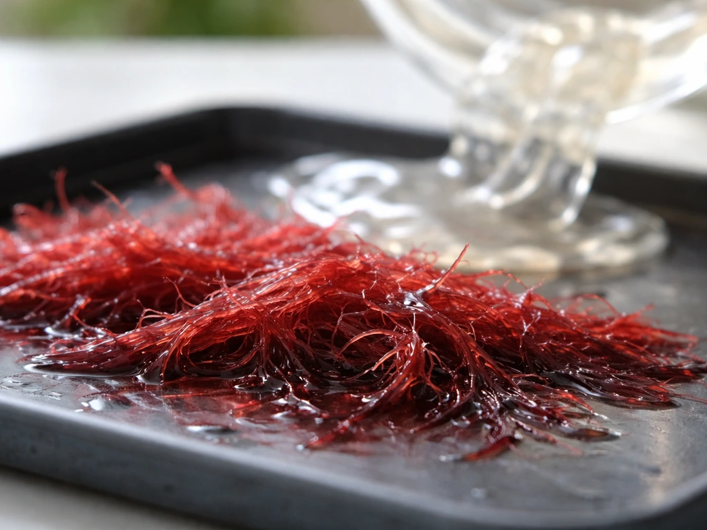
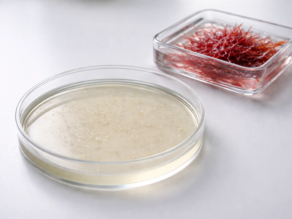
Agar is a natural polysaccharide mixture extracted from certain red algae. It is made up of two fractions: agarose, the high-molecular-weight neutral polymer (above 100,000 Da) that is responsible for most of the gelling, and agaropectin, a smaller sulfated polymer (around 14,000 Da) that contributes very little to gel formation. When you dissolve agar powder in boiling water and let it cool, the agarose chains form a network of hydrogen bonds that traps water and sets into a firm gel.
That gel does not re-melt until the temperature climbs back above roughly 85 °C, but the original setting happens at a much lower 32 to 42 °C. This temperature gap (called hysteresis) is critically useful in the lab, as you will see.
Here is the key point that surprises many students: agar itself is not a nutrient. It is a structural scaffold. Bacteria cannot digest it (almost none produce the enzyme agarase), so the gel sits there unchanged while colonies grow on top of it. If you want bacteria to have food, you add nutrients separately. If you want a blank surface, you leave the agar plain. That separation of structure from nutrition is what makes agar so controllable.
Why agar plates beat every other solid surface
Before agar, microbiologists tried gelatin as a solid medium. Gelatin works at room temperature, but it melts above about 28 °C, which rules it out for incubating most bacteria at their preferred 35 to 37 °C. Worse, many bacteria secrete gelatinase, an enzyme that liquefies the gel and destroys the surface structure you need for colony formation. Agar solved both problems in one move: it stays solid well past any practical incubation temperature, and bacteria almost universally cannot degrade it. That is why agar supplanted gelatin as the foundation of microbiological media and has stayed there for well over a century.
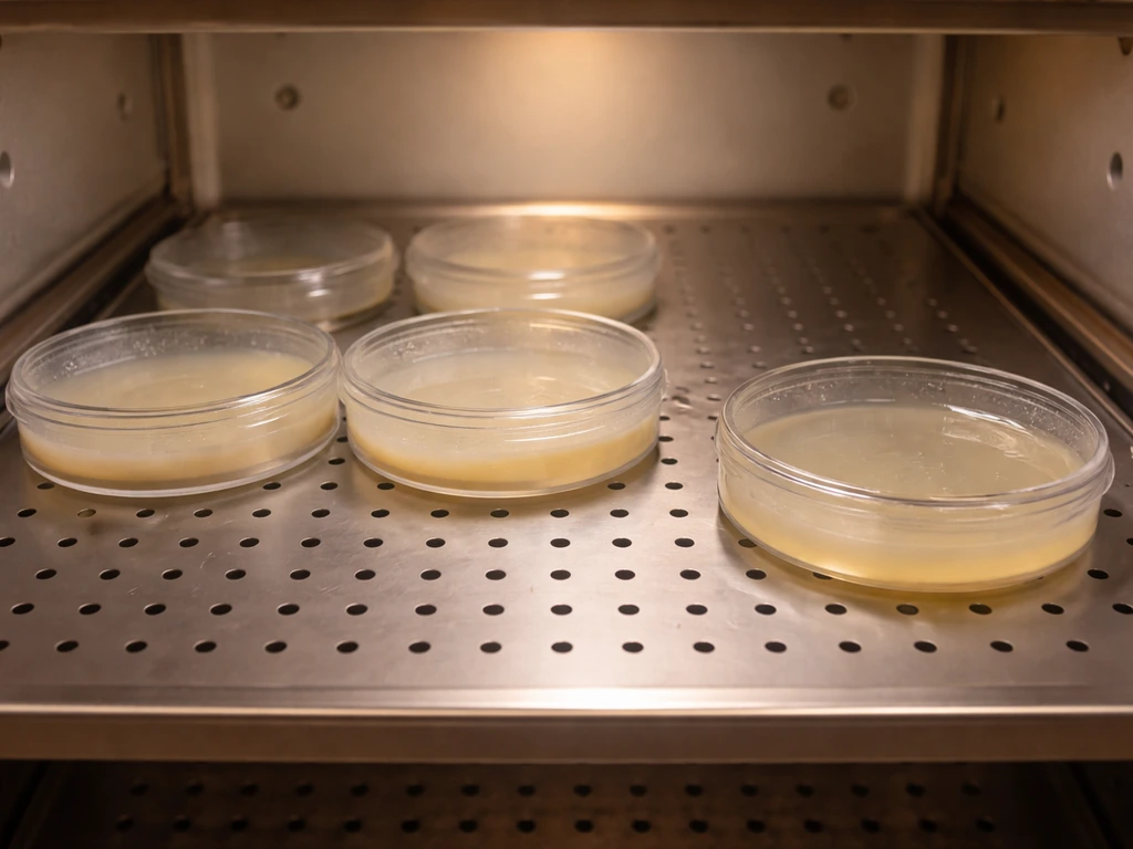
Compared with other solid surfaces you might imagine (filter paper, soil, plastic), agar has additional advantages. It holds a large volume of water in a form that bacteria can access, it can be poured into standard petri dishes to give a uniform, flat surface, it is transparent enough to see colony growth without moving anything, and it is easy to sterilize. You autoclave it at 121 °C under pressure for 15 minutes, killing all contaminants, then pour it while still liquid into sterile plates. Once it sets, you have a ready-to-use sterile surface.
Why nutrient agar is the go-to choice
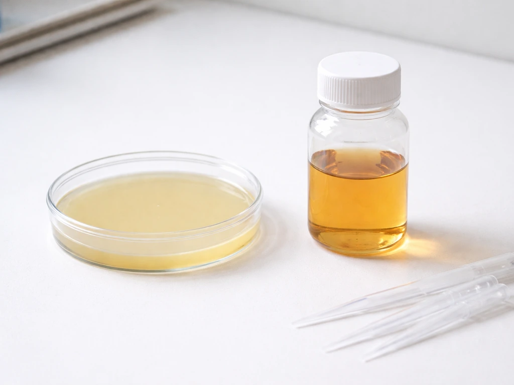
Plain agar gives bacteria a surface but no food. Nutrient agar adds a simple, standardized nutrient base so that most common, non-fastidious bacteria can actually grow. The FDA's Bacteriological Analytical Manual specifies the formula precisely: 3 g of beef extract per liter, 5 g of peptone per liter, and 15 g of agar per liter, made up with distilled water. Beef extract provides vitamins, minerals, and amino acids; peptone supplies nitrogen and additional carbon. Together they support a wide range of bacteria without favoring any particular group.
Compare that with nutrient broth, which has exactly the same nutrient formula but no agar at all. The only difference is that nutrient broth stays liquid, so bacteria grow suspended throughout it rather than on a surface. This comparison makes it obvious that agar's job is purely structural. The nutrients do the feeding; agar just holds everything in place.
When you need selectivity (allowing only certain bacteria to grow) or differentiation (making bacterial groups look different from each other), you swap out or add to the nutrient base while keeping agar as the solidifying agent. [Mannitol salt agar uses high salt to select for halotolerant bacteria and differentiates Staphylococcus aureus by mannitol fermentation](https://bio. libretexts. org/Bookshelves/Microbiology/MicrobiologyLaboratoryManual%28Hartline%29/01%3ALabs/1.
29%3AMannitolSalt_Agar), as reflected in the medium’s reaction. MacConkey agar does this by adding bile salts and lactose to select for Gram-negative bacteria; cetrimide agar adds a disinfectant to target Pseudomonas aeruginosa specifically.
How an agar plate supports the fundamental conditions bacteria need
Bacteria need five things to grow: nutrients, water, an appropriate temperature, a suitable pH, and access to (or protection from) oxygen. An agar plate addresses each of these in a practical way.
Water availability
Agar holds water within its gel matrix and releases it slowly, keeping the surface moist enough for bacterial metabolism without flooding the plate. Bacteria need liquid water to transport nutrients across their membranes; a gel that retains water while still being solid threads that needle perfectly. If the agar dries out and cracks, bacteria will not grow well on the damaged surface, which is why best practices recommend cooling prepared media to around 45 to 50 °C before pouring (to reduce condensation) and not over-drying plates.
Temperature tolerance
This is where agar's physical chemistry really matters. Because the gel does not melt until above 85 °C, you can incubate plates at 37 °C all day without the medium turning back to liquid. Most mesophilic bacteria, which includes the majority studied in classrooms and clinical labs, thrive right in that 35 to 37 °C window. Agar handles that without blinking. You can even prepare thermophilic cultures at higher temperatures without the gel losing its structure, whereas gelatin would have failed long before you reached 37 °C.
pH stability
Agar itself is essentially pH-neutral and does not significantly buffer or alter the pH of the medium. That means you control pH entirely through the nutrient components and any buffers you add. Most bacteria prefer a pH close to 7.0, and standard nutrient agar is formulated to land right there. Because agar does not react with or consume the buffering agents in your medium, the pH you set when you prepare the plates is the pH bacteria experience throughout incubation.
Oxygen access
Colonies growing on the surface of an agar plate are directly exposed to atmospheric oxygen, which suits aerobic bacteria perfectly. Facultative anaerobes (bacteria that can grow with or without oxygen) also thrive on the surface. If you want to culture strict anaerobes, you need to modify the atmosphere around the plate, but the agar medium itself does not impose oxygen restrictions. Deeper in a pour-plate setup, bacteria experience lower oxygen concentrations, which can be exploited deliberately. The point is that agar does not complicate your oxygen control; it leaves that variable entirely in your hands.
What actually happens during incubation: colonies, counting, and isolation
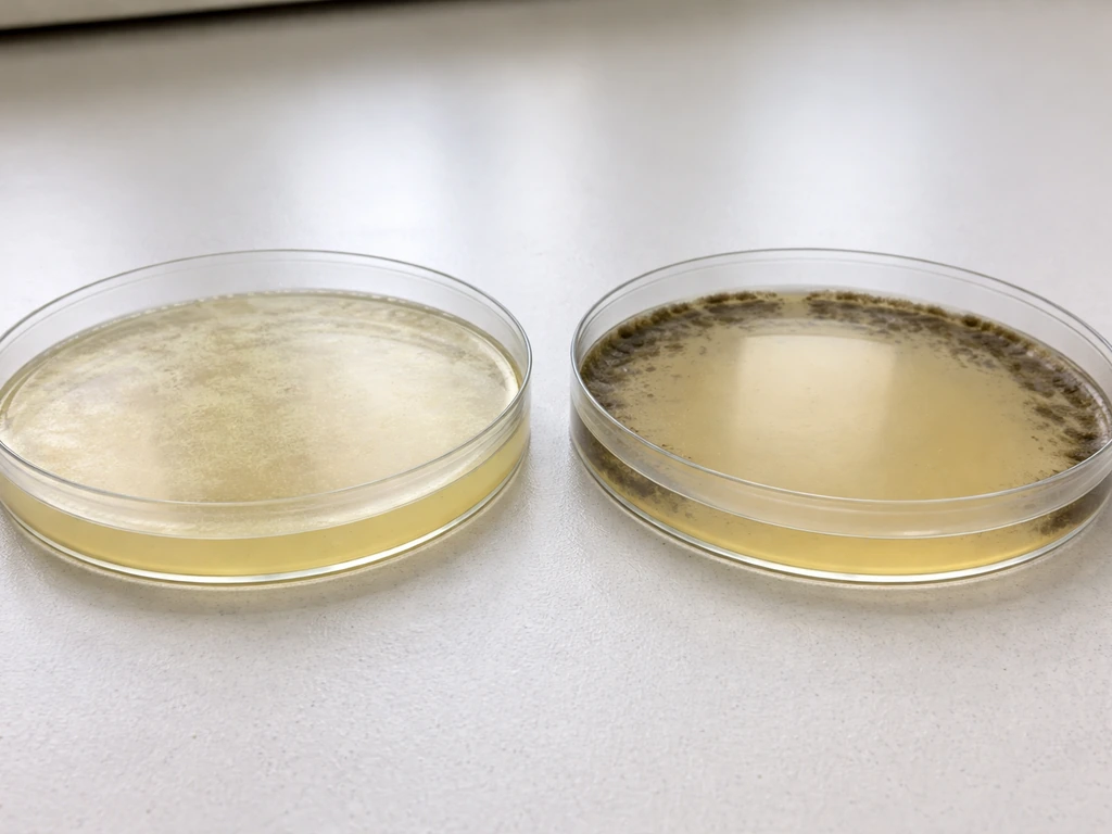
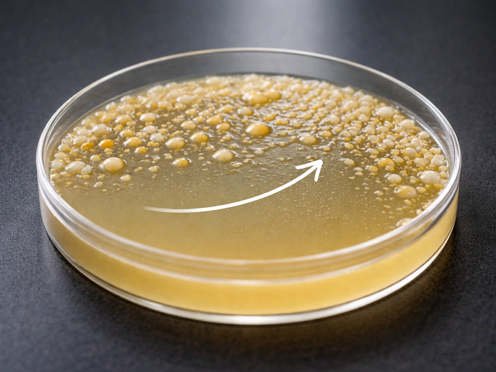
When a single bacterial cell lands on a nutrient agar surface and conditions are right, it divides repeatedly. Because agar is a solid, the daughter cells cannot disperse through the medium the way they would in broth. They pile up in place, forming a visible mound called a colony. Each colony is, in effect, a clone of the original cell. That is why colony morphology (shape, color, texture, size) can tell you a great deal about the organism producing it.
This colony-forming property is what makes agar plates so useful for counting bacteria. In the spread-plate technique, you dilute a sample, spread it evenly across a pre-dried agar surface, and after incubation count the colonies that appear. The standard counting range is 30 to 300 colonies per plate. Below 30, your count is statistically unreliable. Above 300, colonies start crowding each other, inhibiting growth of neighbors and making it impossible to count accurately. Results are reported in colony-forming units per milliliter (CFU/mL) rather than individual cells, because a single colony may have started from a clump of two or three cells rather than one.
Isolation works on the same principle. Streak a mixed sample across an agar plate in a pattern that progressively dilutes the cells, and by the final set of streaks individual cells land far enough apart to grow into separate, well-isolated colonies. You can then pick a single colony and be reasonably confident you are working with a pure culture of one organism, which is the foundation of almost all downstream microbiological work.
Choosing the right agar and avoiding common problems
For general classroom work or routine culturing of non-fastidious bacteria, nutrient agar (beef extract, peptone, agar, water) is the right starting point. Bacteria can grow on potato dextrose agar if the organism’s needs are met, which is why PDA is commonly used for culturing fungi and can also support some bacteria can bacteria grow on potato dextrose agar. It supports a wide range of common organisms without adding complexity. If you are working with E.
coli specifically, you have several options depending on your goal: nutrient agar works fine for basic growth, while MacConkey agar lets you differentiate lactose fermenters in a mixed culture. If you are culturing E. coli specifically, the next question is what agar does E. coli grow on for your intended results.
In liquid media, you generally grow E. coli by supplying the same kinds of nutrients while allowing cells to multiply while suspended in the broth. E. coli can also be grown in liquid media by providing the right nutrients and the right conditions for cells to multiply while suspended E. coli in liquid media. What nutrients E. coli needs to grow depends on the medium, but nutrient agar and related formulations typically supply amino acids, nitrogen, vitamins, and salts E.
If you are looking for a direct breakdown of what nutrients E. coli needs to grow, see what nutrients does e coli need to grow as a related option. In fact, if you want a direct breakdown of what nutrients E. coli needs to grow, see what nutrients does e coli need to grow as a related option. - p25s3: coli specifically. Different agar types suit different questions, and the agar base stays the same while the nutrient or selective components change.
There are a few practical problems that consistently trip up beginners, so it is worth addressing them directly.
- Sterilization: Always autoclave agar media at 121 °C for 15 minutes before pouring. Skipping or shortcutting this step guarantees contamination from environmental organisms landing on unsterilized plates.
- Condensation: Cool melted agar to around 45 to 50 °C before pouring into plates. Pouring it too hot creates a cloud of steam inside the closed lid that condenses into water droplets on the lid surface. Those droplets drip back onto the agar and can cause colonies to smear, making isolation impossible.
- Plate inversion during incubation: Once inoculated, incubate plates upside down (agar side up). This keeps any condensation that forms on the inside of the lid away from the colony surface, preventing droplets from washing cells across the plate and ruining your isolation.
- Pre-warming and drying plates: For spread-plate methods especially, plates should be fully dry and at room temperature before you spread your sample. Moisture on the surface prevents even spreading and leads to crowded, poorly separated colonies.
- Agar concentration: Standard solid media use roughly 1.5% agar by weight (15 g/L as in the FDA formula). Going significantly lower gives a soft, fragile gel; going much higher produces a harder surface that some bacteria find difficult to grow on.
One more thing worth knowing: agar is not universal. Some fungi grow better on specific agar types (Sabouraud's dextrose agar, for instance, which uses a much more acidic pH to suppress bacteria and favor fungal growth). Certain bacteria like Pseudomonas aeruginosa have selective agars designed specifically for their isolation. For example, specialized selective media are used to determine what agar Pseudomonas aeruginosa grows on. Understanding why plain or nutrient agar works gives you the foundation to understand why specialized agars do what they do: they are all built on the same reliable gel matrix, just with different nutrient or inhibitory ingredients layered on top.
| Agar Type | Main Additives Beyond Agar | Best Used For | Selective or Differential? |
|---|---|---|---|
| Nutrient Agar | Beef extract (3 g/L), peptone (5 g/L) | General culturing of non-fastidious bacteria | Neither — general purpose |
| MacConkey Agar | Bile salts, crystal violet, lactose, neutral red | Isolating and differentiating Gram-negative bacteria, including E. coli | Both selective and differential |
| Cetrimide Agar | Cetrimide (antiseptic) | Selective isolation of Pseudomonas aeruginosa | Selective |
| Mannitol Salt Agar | 7.5% NaCl, mannitol, phenol red indicator | Isolating halotolerant staphylococci, especially S. aureus | Both selective and differential |
| Sabouraud's Dextrose Agar | Dextrose (40 g/L), low pH (~5.6) | Culturing fungi while suppressing most bacteria | Selective |
The bottom line is straightforward: agar works because it sits at the intersection of everything bacteria need. It holds water, it stays solid at incubation temperatures, it does not interfere with nutrients or pH, it does not get digested by most organisms, and it provides a flat transparent surface where individual cells become visible colonies you can count, identify, and isolate.
Sigma-Aldrich notes that typical solid media use about 1, 7% agar-agar (or 10, 20% gelatin) to solidify a liquid broth 1–7% agar-agar to solidify a liquid broth. In most cases, you can expect many common bacteria to grow on LB agar because it provides a rich nutrient base that supports broad bacterial growth default solid growth medium.
No other material comes close to checking all those boxes simultaneously, which is why agar has been the default solid growth medium in microbiology for well over 130 years and shows no sign of being replaced.
FAQ
Why doesn’t agar get “eaten” the way gelatin does, and are there exceptions?
Most bacteria do not break down agar because they rarely produce agarase. The key exception is some marine bacteria and certain specialized lab strains that can degrade it, which is why medium choice matters when culturing unusual organisms.
What happens if the agar plate is poured too hot or too cold?
If you pour while the agar is too hot, it can increase condensation and sometimes stress cells on the surface later. If it sets too cold or you wait too long after autoclaving, the agar can start gelling in the bottle, leading to uneven thickness and inconsistent colony spread.
Does agar affect bacterial pH or metabolism?
Agar is largely pH-neutral and does not significantly buffer. However, if you add antibiotics, dyes, or specialized selective agents, those additives can shift effective pH or stress cells, so you still need to verify the full formulation, not just the agar base.
Why does plate drying matter, and how can over-drying change results?
Drying reduces available surface water. That can prevent colonies from forming, shrink colony size, or make spreading look like there is less growth than there really is. Rehydration is not a reliable fix because it can cause smearing or uneven distribution.
Can you store prepared agar plates, and does storage change their performance?
Yes, but quality declines with time. Long storage can reduce moisture, change antibiotic potency (if present), and alter colony morphology. Many labs store plates sealed to limit dehydration and use them within a validated window.
Why do bacteria sometimes fail to grow even on “nutrient agar”?
Growth can fail if the organism is fastidious (needs specific growth factors), if the incubation temperature or oxygen conditions are wrong, if the sample has inhibitory carryover (antibiotics, disinfectants), or if the plate was improperly sterilized or allowed to dry out.
Is “nutrient agar” the same as “LB agar,” and which should I use?
They are not identical formulations, though they overlap in intent (broad, non-selective growth). Use the one that matches your protocol, especially if you are doing quantitative work like CFU counts where slight formulation differences can affect colony yield.
How does agar help with counting bacteria accurately, and why are 30 to 300 colonies special?
That range balances statistics and crowding. Below about 30 colonies, random variation is too large, and above it, neighboring colonies compete for nutrients and space, making individual colonies harder to distinguish and leading to undercounting.
Why does the same bacteria produce different colony shapes on different agar types?
Even with the same gel scaffold, added nutrients and selective or inhibitory components change growth rate, metabolism, and surface interactions. Agar plus an additive can alter colony size, pigmentation, and texture, so colony morphology is not universally comparable across media.
Can agar plates be used for obligate anaerobes without modifying anything?
Not reliably. Agar itself does not block oxygen, so strict anaerobes usually need an anaerobic jar, chamber, or gas-generating system. Without that, oxygen exposure can reduce viability or prevent colony formation.