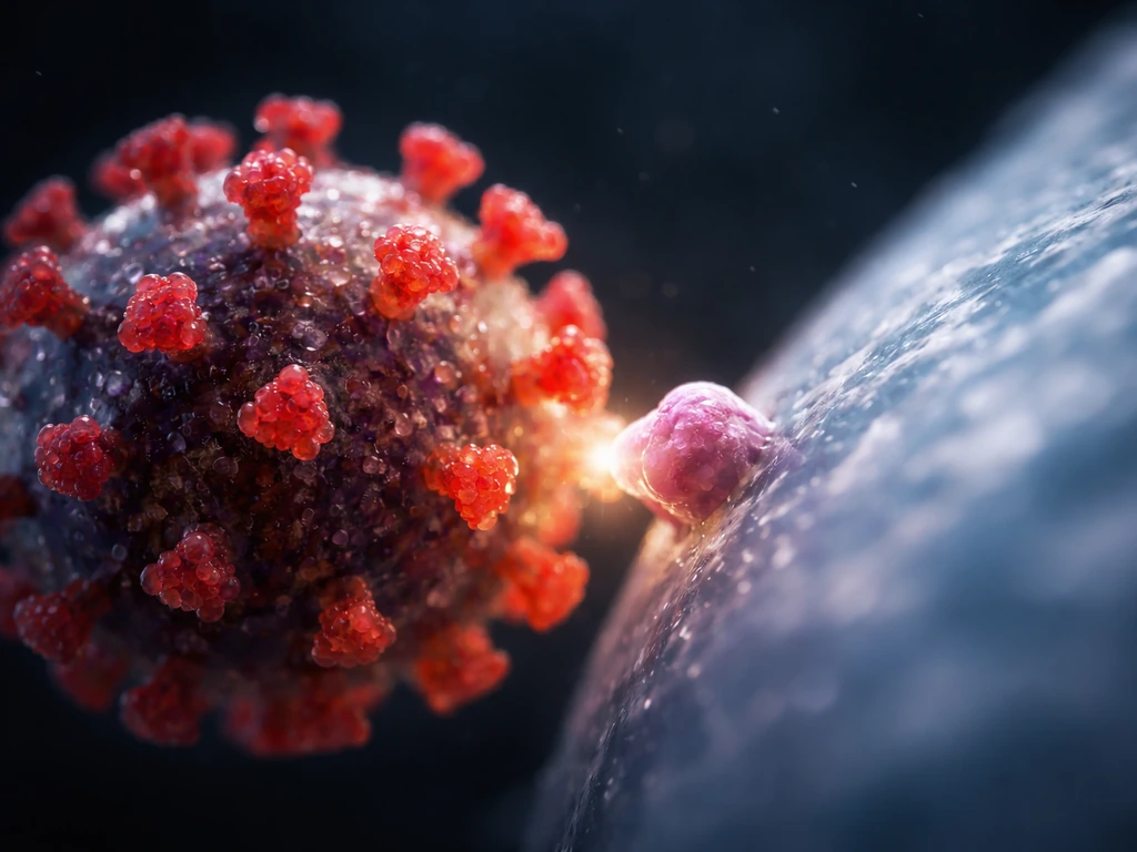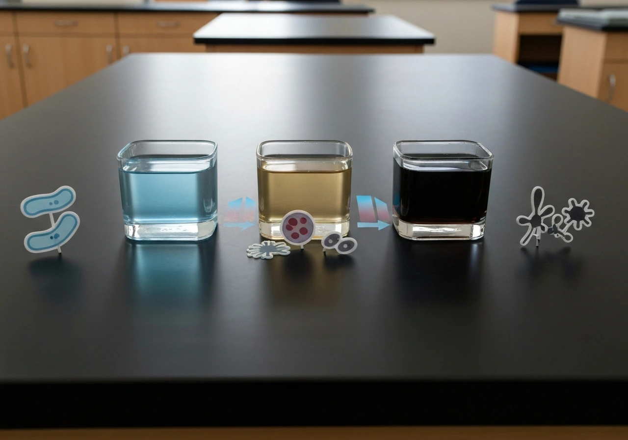
Virologists use embryonated chicken eggs to grow viruses because an egg contains exactly what a virus needs to replicate: living, dividing cells packed into accessible tissues, all wrapped inside a self-contained environment that is easy to control. A virus cannot grow on its own the way bacteria can. It has no metabolism, no energy system, and no way to copy itself without hijacking the molecular machinery of a living host cell. An embryonated egg (a fertilized egg with a developing embryo inside) provides those living host cells in generous supply, at the right temperature, in a sterile sealed package. That combination is hard to beat.
Why Are Embryonated Chicken Eggs Used to Grow Viruses

Marcus Holloway
9 Jul 2026
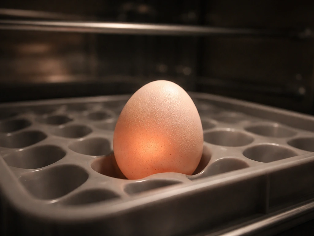
What "growing viruses" in an egg actually means
It is easy to picture growing bacteria in a broth and assume viruses work the same way, but they do not. Bacterial growth is essentially fermentation-style multiplication: bacteria consume nutrients, generate energy, and divide on their own. Viral "growth" is something entirely different. When virologists say they are growing a virus in an egg, they mean they are triggering viral replication inside living embryonic cells. The virus particle enters a cell, takes over its replication machinery, produces thousands of new virus copies, and those copies are released into the fluid surrounding the tissue. That fluid is then harvested as the virus-containing product. There is no free-living viral metabolism happening. If the living cells are gone, replication stops completely. This distinction matters because it explains why you cannot just drop a virus into a nutrient broth and incubate it. The "growth medium" for a virus is a living cell, and the egg is a convenient, self-contained package of those cells.
This is also why viruses are genuinely difficult to grow in a laboratory compared to bacteria or fungi. Bacteria and fungi have their own metabolism and can survive and multiply independently given the right chemical environment. Viruses are entirely dependent on a host cell's internal machinery to copy themselves, which narrows the options for where they can be cultured. The embryonated egg solves that problem elegantly.
Why a fertilized, embryonated chicken egg specifically
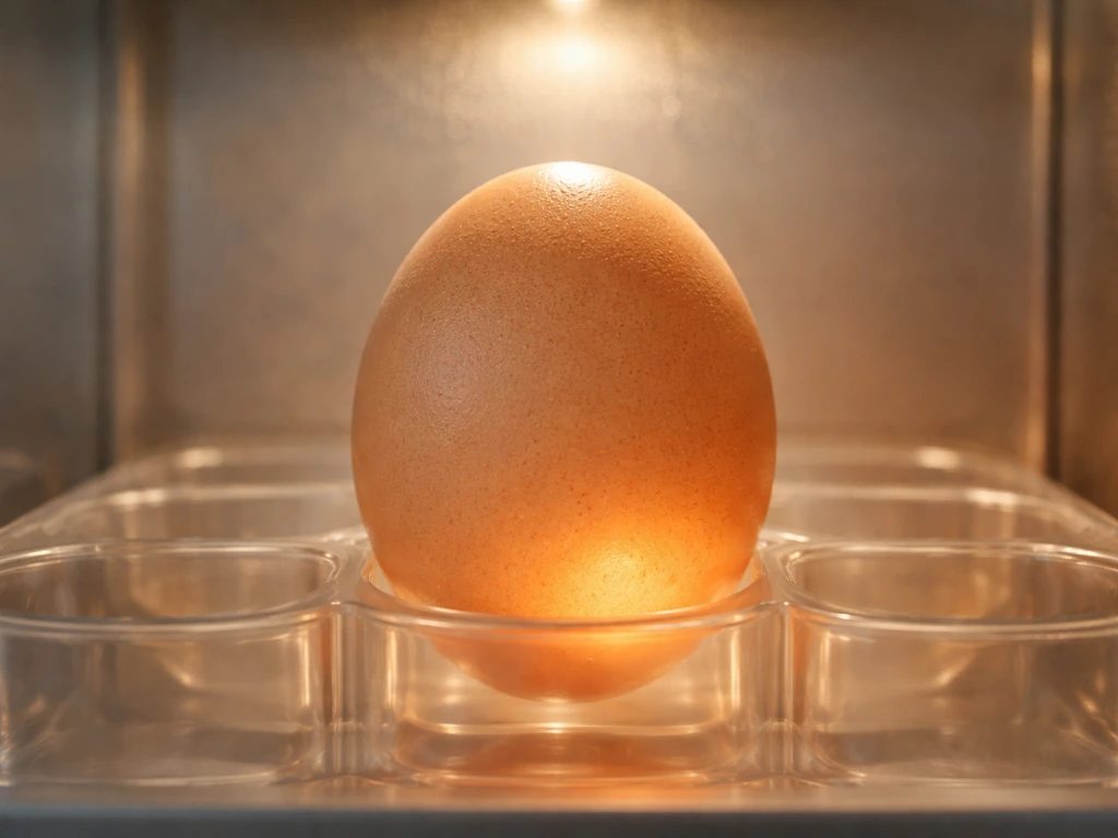
Not just any egg will do. A regular grocery-store egg is unfertilized and contains no living embryo, so there are no dividing cells for a virus to infect. An embryonated chicken egg, fertilized and incubated for several days before use, contains a developing chick embryo surrounded by a set of specialized membranes and fluid-filled compartments. Each of those compartments is lined with different types of actively dividing embryonic cells, and that developmental activity is exactly what makes the egg so useful.
The embryonic tissues are immunologically naive compared to adult tissues. The developing chick has not yet mounted a full immune response, which means many viruses can infect and replicate with fewer obstacles than they would face in an adult animal. The cells are also actively dividing, and rapidly dividing cells tend to be more permissive to viral entry and replication. Many viruses preferentially infect proliferating cells precisely because the cellular machinery they need is running at full speed.
One important misconception to clear up: the virus does not use the yolk or egg white as a nutrient source the way yeast uses sugar in a fermentation vat. The virus uses the cells lining the internal membranes and cavities. The yolk and albumen support the embryo's growth, which keeps those cells alive and dividing, but the virus itself is targeting the cells, not the nutrients directly.
The growth conditions inside the egg that support viral replication
Temperature is one of the most critical variables in any microbial growth system, and viral replication in eggs is no exception. The standard incubation temperature for growing influenza virus in embryonated eggs is 37 degrees Celsius (the same as mammalian core body temperature), with humidity maintained at 55 to 60 percent. Eggs used for influenza work are typically incubated for 10 to 11 days before inoculation to reach the right stage of embryonic development. Research comparing incubation temperatures of 35, 37, and 39 degrees Celsius for avian influenza strains showed measurable differences in viral polymerase activity and final yield, confirming that temperature is not just a background detail but a genuine driver of how well replication proceeds.
Beyond temperature, the egg provides a moisture-rich, chemically balanced internal environment. The allantoic fluid (the fluid in the main working compartment used for influenza production) maintains physiological ion concentrations and pH ranges that keep embryonic cells viable and viral surface proteins functional. The shell and membranes act as a natural barrier, keeping contaminating microorganisms out while maintaining internal humidity. After inoculation, eggs are kept at controlled temperatures for a defined incubation period, then chilled to 4 degrees Celsius before harvest. That chilling step kills the embryo, constricts blood vessels, and reduces blood contamination in the harvested fluid, which matters for getting a clean, high-titer stock.
Timing is also tightly coupled to the biology. The embryonic compartments change as development progresses, and the right stage of development determines which tissues are accessible and most permissive. A 10-day-old embryo has well-developed allantoic and amniotic compartments ideal for influenza work. A 5-day-old embryo might be used for yolk sac inoculations targeting other viruses. Using an egg at the wrong developmental stage is like trying to culture an organism at the wrong temperature: the host cells may simply not be in the right physiological state to support productive infection.
Getting the virus to the right place inside the egg
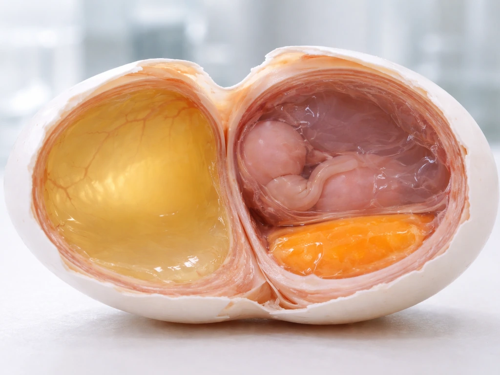
An embryonated egg has several distinct internal compartments, each lined with different tissues, and different viruses need to reach different target cells to replicate productively. The main compartments used in virology work are the allantoic cavity, the amniotic cavity, the blank" rel="noopener noreferrer">chorioallantoic membrane (the CAM), and the yolk sac. Inoculation (injecting the virus into the right compartment through the shell) is the step that puts the virus where it needs to be.
Influenza virus is most commonly inoculated into the allantoic cavity because the cells lining that cavity display the sialic acid receptors that influenza uses to enter cells. The allantoic cavity also produces large volumes of fluid, often 5 to 10 milliliters per egg, which makes it the preferred compartment for generating high-titer stocks. For some influenza strains, the amniotic cavity is preferred because its lining has a slightly different sialic acid linkage pattern that suits certain viral strains better.
Other viruses use other routes. Poxviruses like vaccinia and fowlpox are inoculated onto the chorioallantoic membrane, where they replicate and produce visible lesions called pocks directly on the membrane surface within two to four days. The size and appearance of those pocks can actually be used to differentiate virus strains, which makes the CAM a useful read-out tool as well as a culture site. Turkey coronavirus, by contrast, replicates only in embryonic intestinal tissue and the bursa of Fabricius, meaning only an amniotic inoculation route will deliver the virus to the right cells. This tissue specificity, called tropism, is a consistent theme in virology: the virus must reach cells that carry the specific surface receptors it recognizes, or nothing happens.
How you actually know the virus grew (it is not always obvious)
One thing that surprises students is that viral replication in an egg does not always produce obvious visible damage to the embryo. Sometimes the embryo dies, which is a clear sign of infection. Virulent Newcastle disease virus, for example, invades beyond the allantoic lining and kills the embryo outright. But many viruses replicate productively without causing visible lesions at all. In those cases, growth is confirmed by harvesting the allantoic fluid and running assays: hemagglutination tests (influenza virus particles can clump red blood cells, which gives a quick positive/negative read), RT-PCR to detect viral RNA, or infectious dose calculations using the Reed-Muench method to determine the EID50 (50 percent egg infectious dose), which quantifies how much infectious virus is present per milliliter of fluid. The key point is that "growth" in this system means amplification of infectious virus particles in the fluid, verified by functional assays, not just a visual inspection of the egg.
The practical advantages that keep eggs in use
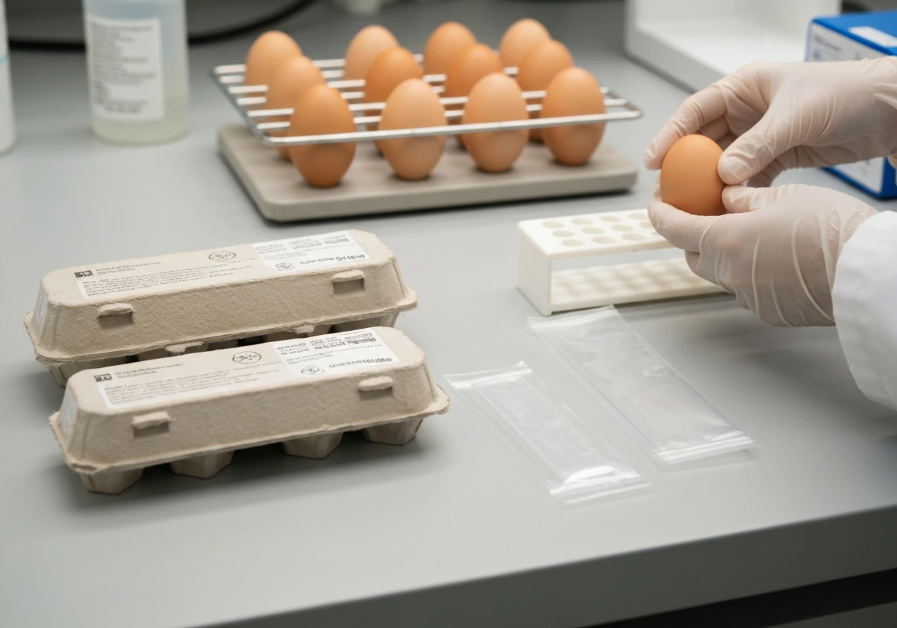
Eggs have been used in virology since the 1930s, and they remain in active use today for several straightforward practical reasons.
- Cost and availability: Embryonated eggs (especially specific pathogen free, or SPF, eggs from controlled flocks) are far cheaper and easier to source than the equipment needed for large-scale cell culture. Each egg is essentially a self-contained bioreactor.
- Volume yield: A single egg can produce 5 to 10 milliliters of allantoic fluid, and large batches of eggs can be processed in parallel. That scalability matters when you need large amounts of virus for vaccine manufacturing or diagnostic stock preparation.
- Built-in sterility: The intact eggshell and membranes create a natural closed system that resists microbial contamination, reducing one of the main headaches of maintaining cell culture lines.
- Established standardization: Protocols for egg-based isolation and titration (including EID50 calculations) are deeply standardized across institutions. WHO, FAO, WOAH, and USDA all have established egg-based methods that allow direct comparisons between labs.
- Sensitivity for certain viruses: Studies comparing egg isolation to cell culture (MDCK and Vero cells) have found eggs perform better for some avian influenza strains and avian paramyxoviruses, making them the preferred choice when sensitivity is the priority.
Eggs vs. other viral culture systems: where each fits
| Culture System | Key Advantage | Key Limitation | Best Used For |
|---|---|---|---|
| Embryonated chicken eggs | High yield, built-in sterility, low cost, standardized protocols | Limited to permissive viruses, requires fertile eggs, ethical considerations for embryo use | Influenza isolation/vaccine production, poxvirus work, avian virus diagnostics |
| Cell culture (e.g., MDCK, Vero) | Broader virus range, defined cell lines, no embryo involved | Higher cost, contamination risk, requires continuous maintenance, some viruses grow poorly | Research on human viruses, viruses not permissive in eggs, mechanistic studies |
| Animal models | Reflects full in vivo biology, immune response included | Highest cost, ethical restrictions, logistical complexity | Pathogenesis studies, vaccine efficacy testing, when no in vitro system is adequate |
The honest answer to which system wins is: it depends on the virus. Eggs are the go-to for influenza, Newcastle disease virus, and certain poxviruses. Cell culture (particularly MDCK cells for influenza) is preferred in many modern research and clinical diagnostic settings because it is faster, more flexible, and works for a wider range of viruses. Animal models are reserved for questions that cannot be answered any other way. Real virology labs often use all three depending on what they are trying to find out.
Limitations and why eggs are not always the answer
Eggs are not universally permissive. These constraints explain why some viruses are difficult to grow in a lab even when researchers have access to standard viral culture tools. Some viruses simply do not replicate in chicken embryo tissues because the cells do not carry the right surface receptors or the viral tropism does not match any available embryonic tissue. SARS-CoV, for example, was experimentally tested in embryonated eggs (both allantoic and yolk sac routes) with limited success, illustrating that the technique has real boundaries. Additionally, prolonged passage of a virus through eggs can introduce mutations, particularly in the receptor-binding sites of influenza hemagglutinin, which is a known concern in vaccine manufacturing because the egg-grown virus can differ slightly from the strains circulating in humans.
There are also practical safety and ethics considerations. Working with embryonated eggs at biosafety level 2 or 3 (depending on the virus) requires proper containment, as the eggs contain actively replicating infectious virus. Researchers working with highly pathogenic avian influenza in eggs, for instance, operate under strict biosafety conditions. From an ethical standpoint, embryonated eggs occupy a middle ground: the developing embryo is not an adult animal, but many institutions consider its welfare in research oversight frameworks, particularly at later stages of development.
A subtler limitation is the accumulation of defective viral genomes during egg passage. Research comparing egg-grown and cell culture-grown virus stocks has found differences in the proportion of defective interfering particles, which can affect experimental reproducibility and the biological properties of the resulting stock. This means that even when eggs work well for growing a virus, the stock they produce is not always identical in character to virus grown by other methods, a consideration that matters for research applications even if it is less critical for vaccine manufacturing.
The bigger picture: eggs as a window into viral growth requirements
What makes the embryonated egg story instructive beyond the specific protocol is what it reveals about what viruses fundamentally need to replicate. Temperature, cellular permissiveness, receptor availability, tissue tropism, and a protected internal environment are not unique requirements of egg culture. They are the universal requirements for viral replication wherever it happens, whether in an egg, a flask of MDCK cells, or the respiratory epithelium of a human host. The egg works because it satisfies those requirements in a controlled, accessible format. Understanding why eggs are used is essentially a case study in the broader principle that viral growth conditions must match the biological needs of both the virus and the host cell it depends on. That connection between the egg system and the fundamental biology of how viruses survive and grow is what makes this topic worth understanding deeply, not just as a technique, but as an illustration of what drives microbial replication at the cellular level.
FAQ
Why can’t viruses just be grown in a nutrient broth the way bacteria are?
Because viruses do not have their own metabolism or energy supply, they cannot reproduce without hijacking host cell machinery. In practice, even if you provide nutrients, nothing will produce new infectious virions unless you also supply living, permissive cells (for example, embryonic cells in an egg).
What exactly makes an embryonated egg different from a regular grocery-store egg?
A grocery-store egg is typically unfertilized, so there are no dividing embryonic cells to support viral replication. An embryonated egg has a developing embryo and specialized membrane-lined tissues, which provides both the host cells and the stage-specific cellular state viruses need.
Do viruses in an egg grow by using yolk or egg white as food?
No. The viral target is the living cells in the internal compartments, not the yolk or albumen as a nutrient source. Those fluids primarily help maintain the embryo and the membranes, keeping the permissive cells alive long enough for replication.
Why is the incubation temperature so tightly controlled for egg-grown viruses?
Temperature shifts how quickly host cells divide and how efficiently viral proteins and polymerases function. Even a few degrees can change polymerase activity and final yield, so labs use calibrated incubators with controlled humidity rather than relying on room-temperature conditions.
Why does the stage of embryo development matter?
Different compartments and cell types mature over time, and the availability of permissive targets changes with development. Using an egg that is too young or too old can mean the virus reaches cells that lack the right receptors or are not in a productive physiological state.
Why do influenza labs often use the allantoic cavity instead of the yolk sac?
For many influenza strains, allantoic lining cells present the receptor linkages influenza recognizes, and the allantoic cavity produces large volumes of harvestable fluid. That combination helps labs obtain higher-titer stocks efficiently.
How do researchers confirm viral growth if the egg shows no visible signs of disease?
They typically harvest the relevant fluid and test for infectious virus using functional assays such as hemagglutination, RT-PCR for viral RNA, or EID50 calculations (Reed-Muench) to quantify infectious dose per milliliter.
Can any virus be grown in embryonated eggs if you try hard enough?
No, success depends on tropism and receptor compatibility. If the virus does not match available embryonic tissues or the needed receptors are absent in the chosen compartment, replication will be limited or absent even with correct timing and temperature.
Why might egg-grown virus behave differently from the same virus grown in cell culture?
Egg passage can select for variants that perform better in that environment, and it can also change the mix of particles, including more defective interfering genomes. The result can affect experimental reproducibility, depending on the study goal.
Is growing viruses in eggs always safer or ethically simpler than other methods?
Not necessarily. Egg work still involves live replicating infectious material and requires appropriate biosafety containment for the specific virus. Ethically, the developing embryo adds oversight considerations, and many institutions treat welfare requirements differently depending on developmental stage and protocol.