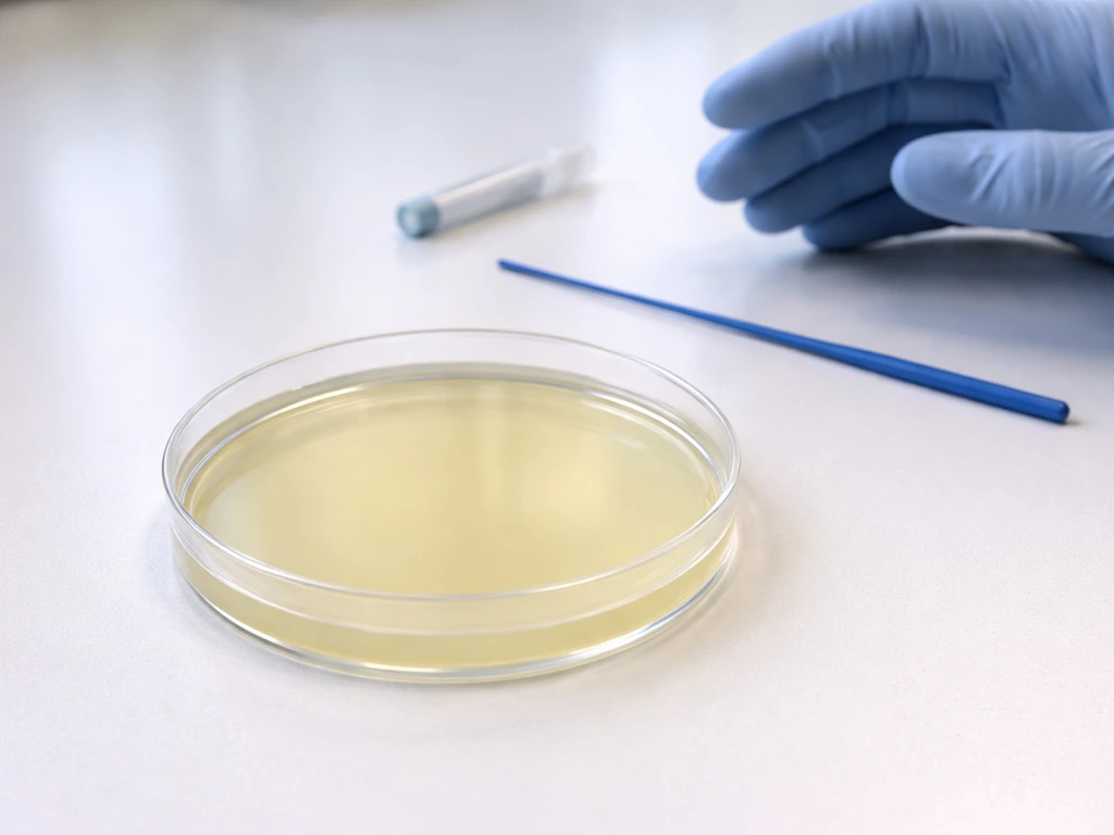
No, you cannot grow viruses on a nutrient agar plate, even one packed with every nutrient a microorganism could want. Viruses are obligate intracellular parasites, meaning they can only replicate inside living host cells. A nutrient agar plate has no living cells, so a virus placed on it simply sits there inert. It does not feed, divide, or produce more copies of itself. The one partial exception worth knowing about is the bacteriophage plaque assay, where agar is used as a scaffold, but the virus is still only replicating inside bacteria embedded in that agar, not in the agar medium itself.
Can You Grow Viruses on an Agar Plate with Nutrients?

Marcus Holloway
16 Jul 2026
Why viruses are host-dependent: the fundamental biology
To understand why agar does not work for viruses, you first need to understand what a virus actually is. A virus is essentially a package of genetic material (DNA or RNA) wrapped in a protein coat called a capsid, sometimes with an outer lipid envelope. It carries no ribosomes, no mitochondria, and no metabolic machinery of its own. It cannot generate energy, it cannot synthesize proteins on its own, and it cannot copy its genome without borrowing the host cell's entire molecular toolkit.
Think of it this way: a virus is like a USB drive with instructions on it. The drive itself cannot do anything until you plug it into a computer with a processor, memory, and software. The host cell is that computer. When a virus infects a permissive host cell, it hijacks the cell's ribosomes to translate its proteins, uses the cell's nucleotides to copy its genome, and exploits the cell's energy supply (ATP) for every step of the process. Without a living, metabolically active host cell, none of this happens. Agar, even nutrient agar enriched with proteins and sugars, provides no living cells, so it provides nothing a virus can actually use.
How viruses differ from bacteria and fungi when it comes to 'growth'
When we talk about growing bacteria or fungi on agar, we mean those organisms are metabolizing nutrients from the medium, producing energy, and dividing on their own. A bacterial cell has everything it needs to multiply in a nutrient-rich environment: enzymes, ribosomes, a cell membrane for energy generation, and the genetic instructions to run all of it. That is why a streak of E. coli on a nutrient agar plate produces visible colonies overnight. Fungi do the same thing with their own metabolic machinery. Growth in both cases means independent cell division fueled by external nutrients.
Viruses do not 'grow' in this sense at all. They replicate, and replication requires a host. The word 'replication' rather than 'growth' or 'division' is deliberate in microbiology because the virus does not split in two like a bacterium does. Instead, it disassembles inside the host cell, directs the host's machinery to produce hundreds of copies of viral components, and those components self-assemble into new viral particles (virions) that burst out to infect neighboring cells. That entire process is cell-dependent at every step.
| Feature | Bacteria | Fungi | Viruses |
|---|---|---|---|
| Living cells | Yes | Yes | No (acellular) |
| Own metabolic machinery | Yes | Yes | No |
| Can replicate on nutrient agar | Yes | Yes | No |
| Requires living host cells | No | No | Yes (obligate) |
| Ribosomes | Yes (70S) | Yes (80S) | None |
| Energy production | Independent | Independent | None, uses host ATP |
| Size range (approximate) | 0.5–5 µm | 2–100+ µm | 20–300 nm |
Where agar does show up in virology: bacteriophage plaque assays
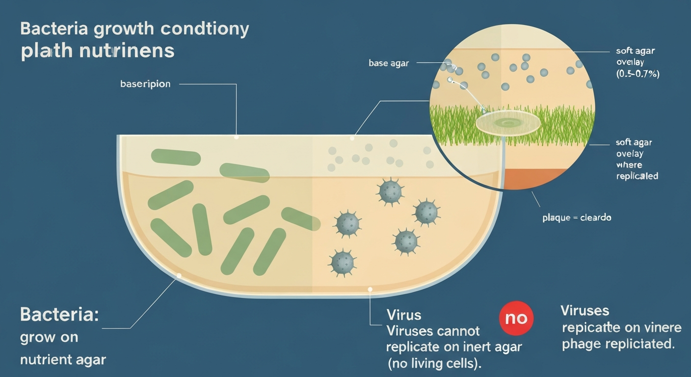
Here is where things get genuinely interesting, and where a lot of students get confused. Agar absolutely appears in virology labs, but its role is not to nourish the virus. It is used as a physical support medium for the living bacterial host cells that the virus actually infects. The technique is called the double agar overlay plaque assay, sometimes called the soft-agar overlay or top-agar method, and it is a foundational tool for studying bacteriophages (viruses that infect bacteria).
How the double agar overlay method works
The assay uses two layers of agar. The bottom layer is a standard hard agar base, typically around 1.5% agar, which acts as a firm foundation. The top layer, called the soft agar or overlay agar, is made with a much lower agar concentration, roughly 0.5 to 0.7% (w/v), which keeps it semi-liquid when warm. You mix your bacteriophage sample and a dense suspension of susceptible host bacteria together into this warm (but not hot) soft agar, then pour it onto the hard base plate and let it solidify. The result is an even lawn of bacteria trapped throughout the soft agar layer.
When the plate incubates (at the host bacterium's growth temperature, such as 37°C for many E. coli phage systems), the bacteria multiply and form a dense, visible lawn across the plate. Wherever a single phage particle landed among the bacteria, it infects, replicates, and lyses nearby cells, then spreads to neighbors. Over several hours, this creates a clearing in the otherwise opaque bacterial lawn. That clearing is a plaque, and each plaque originated from a single infectious viral particle.
- Prepare a hard agar base plate (approximately 1.5% agar in nutrient or LB broth) and let it solidify.
- Serially dilute the phage sample to ensure countable plaque numbers (ideally 20–200 plaques per plate).
- Mix a known volume of the diluted phage with a dense suspension of susceptible host bacteria in molten soft agar (0.5–0.7% agar) held at roughly 45–48°C.
- Pour the soft-agar mixture onto the base plate and swirl gently to distribute evenly; allow to solidify at room temperature for 5–10 minutes.
- Incubate at the host's optimal growth temperature (e.g., 37°C for many E. coli strains) for 8–18 hours.
- Count plaques and calculate the titer using: PFU/mL = (number of plaques × dilution factor) / volume plated in mL.
The resulting number is expressed in plaque-forming units per milliliter (PFU/mL). This is the standard way to quantify how many infectious phage particles are in your sample. Notice that every step relies on the bacteria being alive and actively growing. The agar is simply the scaffolding. Variants of this technique, including spot tests (drop assays) and tube-free overlays, exist to increase throughput in research settings, but they all work on the same principle: phage replicates in bacteria, not in agar. Variants such as spot/drop tests and tube-free overlays have been described to increase throughput and reduce materials; see Streamlining standard bacteriophage methods for higher throughput (PMC methods paper) for validated protocols and guidance on controls and calibration.
Why agar appears in virology but does not support viral replication on its own
It is worth being really precise here because this is a persistent source of confusion. When a virology paper describes 'plating a virus on agar,' what is actually happening is that the virus is being plated alongside living bacterial hosts on agar. The agar is inert from the virus's perspective. It provides no nucleotides, no ribosomes, no energy, and no cell membrane for the virus to exploit. If you streaked a purified viral sample onto a plain nutrient agar plate with no host cells and incubated it under ideal conditions, you would see nothing. No plaques, no colonies, no turbidity. The virus would remain biologically inactive until it encountered a permissive living cell.
The same logic applies to enriched media like blood agar or chocolate agar. These media are enriched with mammalian blood or heat-lysed blood to support fastidious bacteria, but the cells in those preparations are lysed and dead, not metabolically active. A mammalian virus would not replicate in them either. This is a useful distinction to draw because students sometimes assume that 'richer' media should support more types of organisms. For bacteria, richer media often does help fastidious species grow. For viruses, the type of medium is entirely irrelevant since no medium, however nutrient-dense, replaces a living permissive cell.
How eukaryotic viruses are actually cultured in the lab
For viruses that infect animal, plant, or fungal cells (called eukaryotic viruses), researchers use three main approaches: cell culture, embryonated eggs, and animal models. Each method provides living host cells in a controlled setting.
Cell culture: monolayers and suspension cultures
Most eukaryotic virus research today relies on cell culture, sometimes called tissue culture. Scientists grow lines of animal or human cells in flasks or dishes filled with a liquid growth medium supplemented with serum and salts. Common examples include MDCK (Madin-Darby Canine Kidney) cells for influenza viruses and Vero cells (from African green monkey kidneys) for a wide range of pathogens including SARS-CoV-2. These cells attach to the plastic surface of the flask and form a monolayer, a single-cell-thick sheet. When virus is added, it infects the monolayer, and you can observe cytopathic effects (CPE), the visible cell damage and death that virus infection causes, under a microscope. A liquid-medium equivalent of the plaque assay can be performed here too: after adding agar overlay to an infected cell monolayer, plaques of dead or cleared cells become visible, and PFU/mL can be calculated the same way.
Embryonated chicken eggs
Embryonated chicken eggs remain a workhorse for influenza virus propagation and vaccine production. A fertilized egg that has been incubated for about 10 days contains a living embryo surrounded by distinct fluid-filled compartments, including the allantoic cavity, each lined with different cell types. A researcher injects virus into the appropriate compartment (usually allantoic for influenza), reseals the egg, and incubates it for 48–72 hours. The virus replicates inside the embryo's living tissues. Virus yield is recovered from the fluid and quantified using hemagglutination assays or egg infectious dose 50 (EID50) calculations. The WHO manual on animal influenza diagnosis describes these protocols in detail for reference laboratory work.
Animal models
For viruses that do not replicate well in standard cell lines, or for pathogenesis studies, researchers use live animal models such as mice, ferrets, or non-human primates. This approach provides the full complexity of a living immune system and tissue environment, which some viruses require in order to replicate efficiently. Animal work is subject to strict ethical oversight and institutional approval, and it is the most resource-intensive of the three approaches.
What plaques actually tell you (and common misinterpretations)
Plaques are a powerful readout, but they are easy to misinterpret. Each plaque represents one successful infection event initiated by a single infectious viral particle, so the plaque count gives you the number of infectious units in your sample, not the total number of viral particles. These two numbers are often very different. A sample can contain many physically intact but non-infectious virions (damaged capsids, defective genomes) that show up in electron microscopy counts or nucleic acid measurements but contribute zero plaques. The ratio of total particles to infectious particles is called the particle-to-PFU ratio, and in some virus preparations it can be as high as 100:1 or more.
Plaque morphology also tells you something useful. Large, clear plaques often indicate a fast-lysing (virulent) phage. Small or turbid plaques can indicate temperate phages that sometimes integrate into the host chromosome rather than always killing the cell. Plaque size is influenced by incubation time, temperature, agar concentration, and the diffusion rate of the phage, so comparing plaque sizes across experiments requires careful standardization. A common student mistake is to assume that more or larger plaques always mean more virus; in fact, a single dilution series counted at the right dilution (where individual plaques are distinct and countable, ideally 20 to 200 per plate) gives the most reliable titer.
Biosafety: what you can and cannot do in a teaching lab
Working with pathogenic viruses requires biosafety level 2 (BSL-2) containment at minimum, and some respiratory viruses such as SARS-CoV-2 or H5N1 influenza require BSL-3 facilities with specialized ventilation, protective equipment, and strict access controls. The CDC/NIH Biosafety in Microbiological and Biomedical Laboratories (BMBL, 6th edition) and the WHO Laboratory Biosafety Manual (4th edition) are the authoritative references for these requirements. WHO Laboratory Biosafety Manual, 4th Edition (WHO) provides detailed guidance that laboratory propagation and isolation of viruses must follow risk assessment and appropriate containment. No student or teaching lab should attempt to culture pathogenic eukaryotic viruses outside of properly equipped and licensed facilities.
That said, teaching labs can and do work with bacteriophages safely. Programs like HHMI SEA-PHAGES have shown that students can discover, isolate, and characterize novel phages infecting non-pathogenic bacterial hosts (such as Mycobacterium smegmatis) in a standard BSL-1 teaching laboratory setting with appropriate institutional oversight. The American Society for Microbiology (ASM) guidelines for biosafety in teaching laboratories provide a framework for exactly this kind of work, and they are a practical starting point for any educator designing a phage-based curriculum.
When plates show no growth at all: diagnosing common failures
Understanding why a plate fails to show growth is a critical skill that connects across many areas of microbiology, from clinical diagnostics to classroom experiments. The reasons are almost always one of a handful of categories, and working through them systematically saves enormous amounts of time.
Wrong medium for the organism
Nutrient agar supports a broad range of bacteria, but fastidious organisms (those with complex nutritional requirements) may fail to grow on it entirely. A classic example is Lactobacillus species, which grow poorly or not at all on plain nutrient agar because they require specific amino acids, vitamins, and fermentable carbohydrates that nutrient agar does not provide in adequate amounts. MRS (de Man, Rogosa, and Sharpe) agar was specifically formulated to support Lactobacillus growth. Using the correct medium for your target organism is the single most important variable to confirm before troubleshooting anything else.
Incorrect incubation conditions (temperature, atmosphere, time)
Temperature and atmosphere mismatches are extremely common causes of no-growth results. Most lab bacteria are incubated at 37°C (human body temperature), but environmental isolates or food-associated bacteria may have very different optima. Atmosphere matters just as much: obligate anaerobes will not grow in air, and microaerophilic bacteria like many Lactobacillus strains grow best at reduced oxygen concentrations (typically 5% CO2, or in an anaerobic jar). For specifics on optimal incubation temperatures for Lactobacillus species, see what temperature does Lactobacillus grow. For details on preferred temperature, pH, and oxygen conditions, see lactobacilli grow best under. Incubating a microaerophile on an open bench overnight in full air can produce no growth even on a perfectly formulated medium.
Antibiotic selection failures: the LB/ampicillin case
If you are doing a transformation experiment and plating on LB/ampicillin agar to select for bacteria that took up a plasmid, several things can cause total plate failure. Ampicillin degrades over time, especially at room temperature or after freeze-thaw cycles, so old or improperly stored plates will not maintain selection pressure. The standard working concentration is approximately 100 µg/mL of ampicillin (or its more stable analog carbenicillin), and the concentration must be correct in the molten agar before the plates are poured. Transformation efficiency also matters: if the heat shock step was too long, the competent cells were left on ice for too long after heat shock, or the recovery period in SOC medium was skipped, the number of successfully transformed cells may be too low to produce visible colonies. Kanamycin selection (typically 50 µg/mL) has different stability characteristics and similar considerations. Always check the plasmid datasheet to confirm the correct antibiotic and concentration.
Clinical specimens: urine cultures and pre-analytic variables
A 'no growth' result on a clinical urine culture does not always mean the urine is sterile or that the patient has no infection. If a report states that a urine culture did not grow bacteria, consult this detailed explanation of common causes and recommended next steps. Pre-analytic factors are responsible for a significant proportion of false negatives in clinical microbiology. If the specimen was not refrigerated or preserved after collection, organisms can die off or fail to remain viable long enough to grow on the plate. Prior antibiotic use by the patient is another major confounder: even a partial course of antibiotics can suppress bacterial counts below the threshold the lab's plating method can detect. Fastidious urinary pathogens may also fail to grow on the standard culture media used in many clinical labs unless special media or extended incubation is specifically requested. The CDC and IDSA/ASM both recommend prompt processing (within 2 hours at room temperature, or refrigeration up to 24 hours if delays are unavoidable) and the use of preservative transport tubes when timing is uncertain.
Contamination masking true results
Contamination can paradoxically cause apparent 'no growth' of the target organism by overgrowth with contaminants, especially on non-selective media. A heavily contaminated urine sample showing more than three distinct colony morphologies typically indicates contamination from skin flora during collection rather than a true polymicrobial infection. In research settings, contamination of a transformation plate with antibiotic-resistant environmental bacteria can produce false-positive colonies, while contamination of a phage plaque assay with a competing phage strain can distort plaque counts and morphology.
| Problem | Likely cause | How to diagnose / fix |
|---|---|---|
| No growth on nutrient agar (bacterial) | Wrong medium for fastidious organism | Switch to organism-specific medium (e.g., MRS for Lactobacillus, blood agar for Streptococcus) |
| No growth at correct temperature | Wrong atmosphere (O2 level) | Check oxygen requirements; use anaerobic jar or CO2 incubator as needed |
| No colonies on LB/amp plate post-transformation | Degraded ampicillin or wrong concentration | Use fresh plates; confirm 100 µg/mL ampicillin or switch to carbenicillin |
| No colonies on LB/amp plate post-transformation | Poor transformation efficiency | Optimize heat shock; include SOC recovery; check competent cell quality |
| No growth on urine culture | Prior antibiotics or delayed transport | Note antibiotic history; refrigerate or use preservative transport tube |
| No plaques in phage assay | Low phage titer or wrong host strain | Confirm host strain permissiveness; re-dilute sample; check agar concentration and temperature |
| Overgrowth / contamination | Non-selective medium or improper technique | Use selective medium; reinforce aseptic technique; repeat with fresh specimen |
Putting it all together: the core principle
The reason viruses cannot grow on nutrient agar is not a quirk of chemistry or a limitation of the medium's formulation. It is a fundamental consequence of what viruses are: molecular instructions that can only be executed inside a living cell. Every virology technique that involves agar, from phage plaque assays to monolayer overlays, works by providing those living cells as the actual substrate for viral replication. The agar itself is just scaffolding. Understanding this distinction sharply clarifies a huge amount of confusion students encounter when first reading virology protocols, and it reinforces a broader principle that applies across microbiology: the growth requirements of any microorganism are inseparable from its biological nature. Knowing what a microorganism is tells you most of what you need to know about how and whether it will grow under any given set of conditions.
FAQ
Can you grow viruses on a plain nutrient agar plate with nutrients alone?
No. All true viruses are obligate intracellular parasites that require living host cells (bacterial, animal, plant or fungal) to replicate. Cell‑free nutrient agar lacks the cellular machinery (ribosomes, polymerases, metabolic pathways) that viruses need, so viruses cannot replicate or form colonies on plain agar.
In what specific contexts is agar used in virology?
Agar is used indirectly in virology when a living host is present on the plate. The main context is bacteriophage (phage) work: a susceptible bacterial lawn is grown on agar and a phage sample is applied. Using a soft‑agar (double/overlay) plaque assay, lytic phage produce clear zones called plaques where host bacteria are killed. Agar also supports cell culture plates with extracellular matrices, but eukaryotic viruses are propagated in living cell monolayers, eggs, or animals rather than on nutrient agar alone.
What is a double (soft‑) agar overlay / plaque assay and why does it work?
A double‑layer plaque assay uses a firm agar base plus a thin top layer of low‑percent (typically ~0.5–0.7%) agar mixed with host bacteria and the phage. If phage infect and lyse nearby bacteria, each infectious particle can produce a localized clearing (plaque). Counting plaques at known dilutions yields plaque‑forming units (PFU/mL), a quantitative titer of infectious phage.
Can eukaryotic viruses (animal/plant/human) be cultured on agar plates in the same way as bacteria or phage?
No. Eukaryotic viruses require permissive living host cells: cell cultures (e.g., Vero, MDCK), embryonated eggs (e.g., for many influenza strains), or whole animals. These substrates provide the intracellular environment needed for viral replication. Agar plates without living cells cannot support eukaryotic virus propagation.
What laboratory methods are used to grow or propagate viruses other than phage plaque assays?
Common virology culture methods: 1) Cell culture monolayers or suspension cultures (specific cell lines permissive for the virus); 2) Embryonated chicken eggs (standard for some influenza viruses and vaccine production); 3) Animal models for certain research or diagnostics. All require viable host tissue and appropriate biosafety containment.
Why is it important to distinguish bacteriophage culture from culturing pathogenic viruses on agar?
Phage plaque assays involve growing bacteria on agar and are performed with bacteriophages that infect bacteria, not human pathogens. Plain agar cannot grow pathogenic eukaryotic viruses. Misunderstanding this could lead to unsafe attempts to ‘‘grow’’ human viruses on plates or inappropriate handling of pathogenic agents. Virology work must follow institutional biosafety and legal regulations.